Foundations and Historical Development of Transplantation Immunology
Revised 18 November 2026
Introduction to Module 1
Transplant immunology has evolved considerably over the past several decades, shaped by discoveries in immunology, histocompatibility, transplantation biology and clinical practice. Understanding how the field emerged, how its central concepts developed and how they relate to one another provides an important starting point for exploring the science of transplantation. This module is not intended to revisit the general principles of immunology or provide a comprehensive, mechanistic account of transplant immunology. Instead, its purpose is to orient readers to the field, establish its historical and conceptual foundations, and provide an overview of the discoveries and developments that have shaped modern transplant immunology. This broader perspective prepares readers for the subsequent modules, which explore specific areas, concepts and mechanisms in greater depth.
Conceptual overview and historical timeline
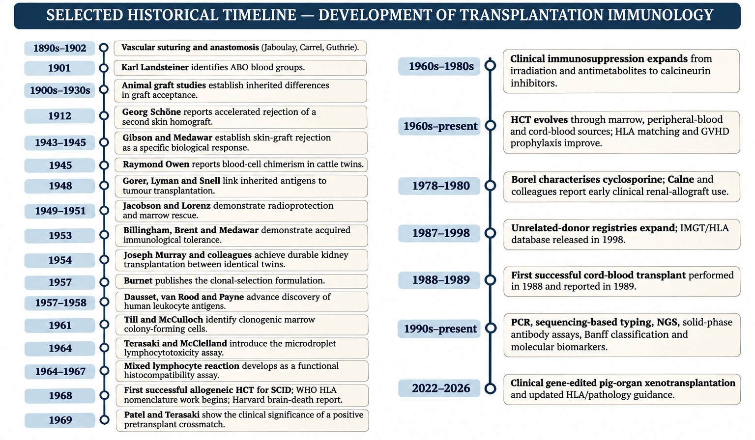
Transplantation immunology emerged when surgeons and experimental biologists recognised that successful tissue transfer required more than restoration of blood flow and anatomical continuity. Grafts could be technically viable yet undergo a reproducible, recipient-directed inflammatory destruction. The decisive shift was to interpret rejection as a specific immune response against genetically determined donor antigens. This insight connected transplantation to fundamental immunology: antigen recognition, clonal expansion, memory, MHC-restricted T-cell function, antibody-mediated injury and immune tolerance.
The historical sequence is not simply a parade of surgical milestones. Vascular anastomosis made organ transplantation technically possible; skin grafts made rejection experimentally observable; inbred strains exposed its genetic basis; H-2 and HLA research identified major histocompatibility systems; tolerance studies demonstrated that alloreactivity could be selectively altered; and histocompatibility testing, immunosuppressive pharmacology and transplantation pathology translated those discoveries into clinical practice. Modern precision transplantation extends the same logic by asking which immune mechanism is active in a particular graft, at what time, in which tissue compartment, and whether intervention can be tailored without broadly eliminating protective immunity.
Landmark discoveries and Nobel recognition
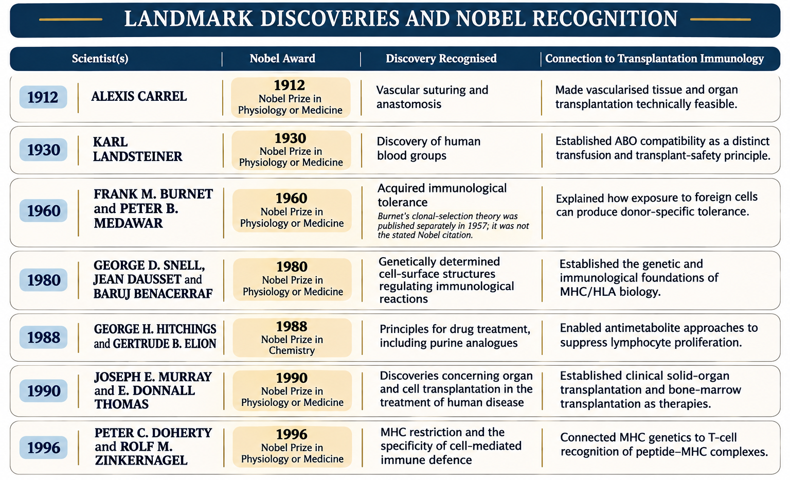
The Nobel Prizes highlighted several discoveries that transformed transplantation from an operative procedure into a discipline grounded in immunogenetics, adaptive immunity and immune modulation. The awards recognised broader scientific contributions; the transplantation relevance shown below is an educational connection, not a claim that each award was given specifically for transplantation.
1.1.1 Origins of Transplantation and Early Experimental Foundations
Transplantation before modern immunology
The idea of replacing damaged tissue with tissue taken from another site or individual is much older than transplantation science. Historical accounts describe reconstructive procedures using local tissue flaps and grafts, including skin and nasal reconstruction. These procedures demonstrate the long-standing surgical aspiration to replace lost tissue, but they should not be confused with the modern transfer of vascularised organs or with a mechanistic understanding of allograft immunity. Most early successes involved autologous tissue, where donor and recipient are the same individual and therefore share the same inherited antigenic constitution. The immunological problem became apparent only when tissue was transferred between genetically different individuals.
The vascular problem and experimental surgery
A major technical barrier was the need to reconnect donor and recipient blood vessels without causing haemorrhage, thrombosis, stenosis or ischaemic necrosis. Work on vascular suturing by surgeons including Mathieu Jaboulay, Alexis Carrel and Charles Guthrie made reproducible vascular anastomosis possible. Carrel’s Nobel lecture describes the experimental development of vascular anastomosis from 1902 onward and its application to vascular and organ transplantation. Carrel’s work was foundational, although the history was collaborative: earlier vascular-suture techniques and Guthrie’s experimental contributions should not be erased by attributing the entire achievement to one individual.
Graft categories and the logic of experimental comparison
Transplantation experiments require explicit definition of the genetic relationship between donor and recipient. An autograft is transferred within one individual. A syngeneic or isograft is transferred between genetically identical individuals, such as inbred animals or monozygotic twins. An allograft (historically, homograft) is transferred between genetically distinct individuals of the same species. A xenograft (historically, heterograft) is transferred between different species. These categories are not merely descriptive: they frame the expected antigenic disparity and therefore the likely immune barriers. Autografts and syngeneic grafts generally provide controls for surgical injury and tissue healing; allografts reveal alloimmune responses; and xenografts introduce additional interspecies barriers, including pre-existing natural antibodies, complement activation and species-specific incompatibilities.
From technical feasibility to biological compatibility
The early lesson was that a graft could be perfused and initially viable yet fail because the recipient’s immune system recognised donor-derived antigens. Conversely, graft survival could occur when genetic disparity was absent or when the relevant immune response was suppressed or tolerised. This distinction separated technical success from biological acceptance. It also established a central experimental principle: surgical variables, organ preservation, vascular injury and immune recognition must be analysed independently, even though they interact in real grafts. The same distinction remains essential when interpreting early graft dysfunction today: not all graft injury is rejection, and not all rejection is explained by a surgical complication.
ABO blood groups and early human transplantation attempts
Karl Landsteiner described the ABO blood-group system in 1901, establishing that naturally occurring antibodies can react with donor erythrocyte antigens. ABO is not an HLA system: ABO carbohydrate antigens are expressed on erythrocytes and, variably, on vascular endothelium and other tissues. In a vascularised organ, pre-existing recipient anti-A or anti-B antibodies can bind graft endothelium, activate complement and cause severe antibody-mediated injury. ABO compatibility therefore became an essential, conceptually separate element of donor-recipient assessment alongside HLA typing and crossmatching.
Why early human kidney attempts failed?
Human kidney transplantation was attempted before effective immunosuppression, reliable organ preservation and modern histocompatibility assessment were available. Voronoy’s 1933 attempt using a deceased-donor kidney is a landmark early operation, but it did not produce durable graft function. Post-war programmes in Europe and North America, including work associated with Küss, Hamburger and Hume, helped refine operative technique and explore temporary graft function, yet rejection, ischaemic injury and the absence of effective immunosuppression prevented reproducible long-term success. Historical “firsts” must therefore distinguish an attempt, a technically completed operation, transient function and durable graft survival.
Non-immunological enablers of clinical transplantation
Transplantation became a sustainable clinical therapy only when non-immunological systems developed alongside immunosuppression. Chronic haemodialysis became more feasible after the development of durable vascular access in 1960, providing a bridge for some patients with kidney failure and a means of supporting patients while transplantation programmes matured. The 1968 Harvard Ad Hoc Committee report proposed irreversible coma as a criterion for death, helping address the ethical and practical conditions for deceased-donor procurement in the era of mechanical ventilation. Organ preservation, cold ischaemia management, retrieval surgery, transport logistics, allocation systems and coordinated donor registries subsequently broadened access. Definitions of death, consent, allocation and organ-procurement policy remain jurisdiction-specific; they should not be taught as a single globally uniform system.
Why the early foundations still matter?
The modern field retains the experimental architecture developed in this era. Isografts and allografts are compared to isolate alloimmune effects; donor and recipient genetics are controlled or measured; graft tissue is sampled over time; and immunological outcomes are related to function and pathology. The historical shift from “can the graft be connected?” to “why is the graft accepted or destroyed?” created transplantation immunology as a distinct discipline. It also foreshadowed the contemporary requirement to integrate surgical, histological, serological, cellular and molecular evidence rather than infer a mechanism from graft function alone.
1.1.2 Early Skin Grafting Experiments and the Discovery of Graft Rejection
Skin as an experimental system
Skin became a powerful model because it is accessible, can be transplanted in defined pieces, and displays visible changes during graft failure. During the Second World War, skin grafting was used in the treatment of severely burned servicemen and civilians. Surgeons observed that autografts were generally retained, whereas skin from unrelated donors often failed after an initial period of apparent take. These clinical observations raised the possibility that graft loss was an active host response rather than simply a failure of surgical technique or wound healing.
The experimental work of Gibson and Medawar
Gibson and Medawar’s 1943 study of human skin homografts, followed by Medawar’s controlled rabbit experiments in 1944 and 1945, provided the experimental basis for interpreting graft rejection as an immune phenomenon. The graft initially vascularised and survived, then became infiltrated by inflammatory cells and underwent progressive tissue destruction. Autografts behaved differently, remaining viable when appropriately performed. This contrast was critical: it showed that graft failure depended on the biological relationship between donor and recipient and could not be explained solely by the trauma of transplantation. Medawar’s work was not the first observation of allograft rejection, but it supplied rigorous experimental analysis and linked the phenomenon to acquired immunity.
First-set and second-set rejection
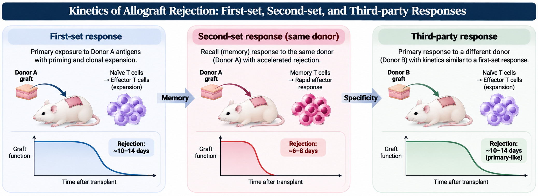
A first allogeneic skin graft commonly undergoes rejection over approximately 10–14 days in classic mouse models, although timing varies with species, strain, antigenic disparity, graft site and experimental conditions. A second graft from the same donor or expressing the same relevant alloantigens is rejected more rapidly. This accelerated second-set response is antigen-specific: a third-party graft with a different antigenic constitution follows a primary-response pattern. Janeway’s describes first-set rejection at roughly 10–13 days and second-set rejection in approximately 6–8 days. The kinetics provide an early demonstration of adaptive immune memory directed against donor alloantigens.
Cellular basis and adoptive-transfer evidence
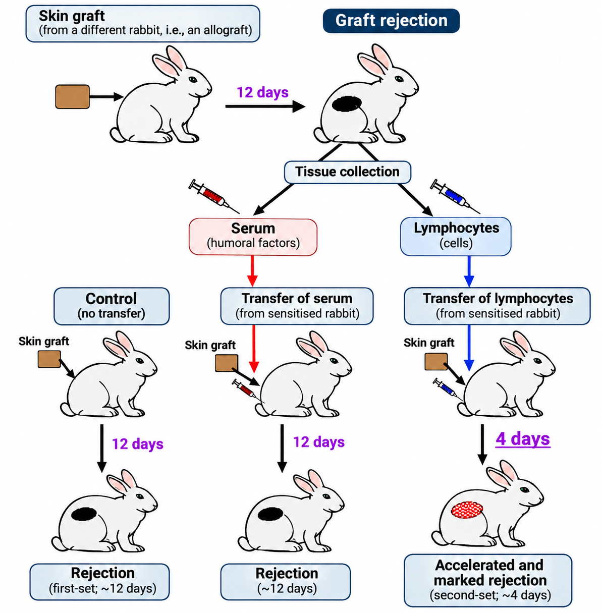
Subsequent experiments established the central role of T lymphocytes. Skin grafts in T-cell-deficient nude mice are not rejected normally, and the capacity to reject can be restored by transfer of normal T cells. T cells from a previously sensitised recipient can transfer accelerated rejection to a naive recipient, directly linking the second-set response to antigen-experienced lymphocytes. Histological studies show dense mononuclear infiltrates and graft-cell injury. These findings place cellular immunity at the centre of classical allograft rejection, while later work established that antibodies, complement, innate leukocytes and endothelial activation also contribute to graft injury.
Specificity, memory and the clinical meaning of sensitisation
The second-set phenomenon anticipated the clinical observation that prior exposure to donor antigens can alter the risk of a subsequent transplant. Pregnancy, blood transfusion and previous transplantation can generate memory B cells, plasma cells and donor-reactive antibodies; cellular memory can also persist after antigen exposure. The immunological response to a graft is therefore not necessarily a first encounter. In clinical histocompatibility, this history is captured by antibody screening, specificity analysis, donor-specific antibody (DSA) assessment and crossmatching. The skin-graft model thus provided more than a historical demonstration of rejection: it introduced the concepts of donor-specific sensitisation and immune memory that underpin modern immunological risk assessment.
Immune privilege: the cornea as a relative model
The cornea illustrates that tissue context modifies the expression of alloimmunity. Its avascularity, relative absence of conventional lymphatic drainage under steady-state conditions, local immunoregulatory mechanisms and specialised ocular microenvironment contribute to relative immune privilege. This does not mean that corneal tissue is invisible to the immune system or that rejection cannot occur. Inflammation, infection, trauma, neovascularisation and disruption of local regulation can increase antigen access and immune priming, raising the risk of graft rejection. Immune privilege is therefore a tissue-specific and conditional state, not absolute immune ignorance.
1.1.3 Discovery of Immunological Specificity and the Genetic Basis of Graft Rejection
From reproducible rejection to a genetic question
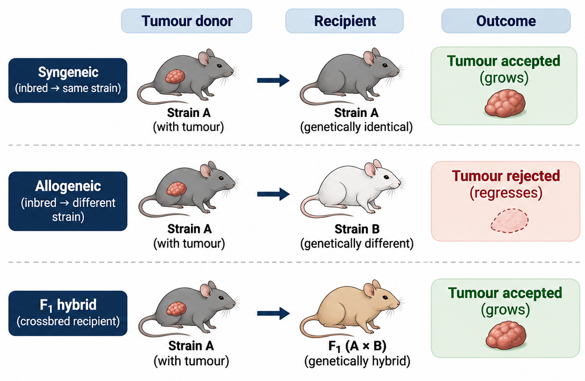
Once allograft rejection was recognised as an immune response, the next question was why a graft from one donor was accepted while a graft from another donor was rejected. Early tumour-transplantation studies in mice had already suggested that susceptibility to transplanted tumours was inherited. Ernest Tyzzer and Clarence Little used crosses between mouse strains to show that tumour acceptance or rejection was strongly influenced by genetic constitution. These experiments did not yet identify the responsible molecules, but they established that graft outcome was not random and could be mapped to inherited determinants.
Gorer, Snell and the antigenic basis of transplantation
Peter Gorer’s work in the 1930s identified serologically detectable antigenic differences among inbred mouse strains and linked one of these antigens to the growth or rejection of transplanted tumours. He showed that animals lacking the relevant antigen rejected tumours expressing it, whereas animals sharing the antigen were more permissive. George Snell subsequently used genetically defined mouse strains to dissect the inheritance of histocompatibility. Gorer, Lyman and Snell’s 1948 work connected a histocompatibility gene with tumour transplantation and helped establish the principle that inherited cell-surface antigens determine important aspects of graft compatibility.
Specificity is not the same as genetic identity
The genetic relationship between donor and recipient predicts, but does not fully define, the immune response. A syngeneic graft is generally accepted because donor and recipient share the relevant inherited antigens. An allograft may be rejected because the recipient recognises polymorphic donor molecules as foreign. However, the strength of the response varies with the number and immunogenicity of mismatched antigens, their expression in the graft, antigen presentation, pre-existing immunity and the inflammatory context. The discovery of major histocompatibility loci did not mean that every other polymorphism was irrelevant; rather, it revealed genetic regions with unusually strong effects on transplantation.
Minor histocompatibility antigens and residual disparity
Even when donor and recipient are matched at major histocompatibility loci, polymorphic proteins encoded elsewhere in the genome can generate peptides that differ between individuals. When these peptides are processed and presented by shared MHC molecules, they can be recognised by T cells as minor histocompatibility antigens. This explains why an HLA-identical sibling graft is not immunologically equivalent to an autograft or an identical-twin graft. It also explains why minor antigens remain relevant in HCT, where donor T cells encounter recipient tissues and can mediate GVHD even when HLA matching is excellent.
The emergence of clonal specificity
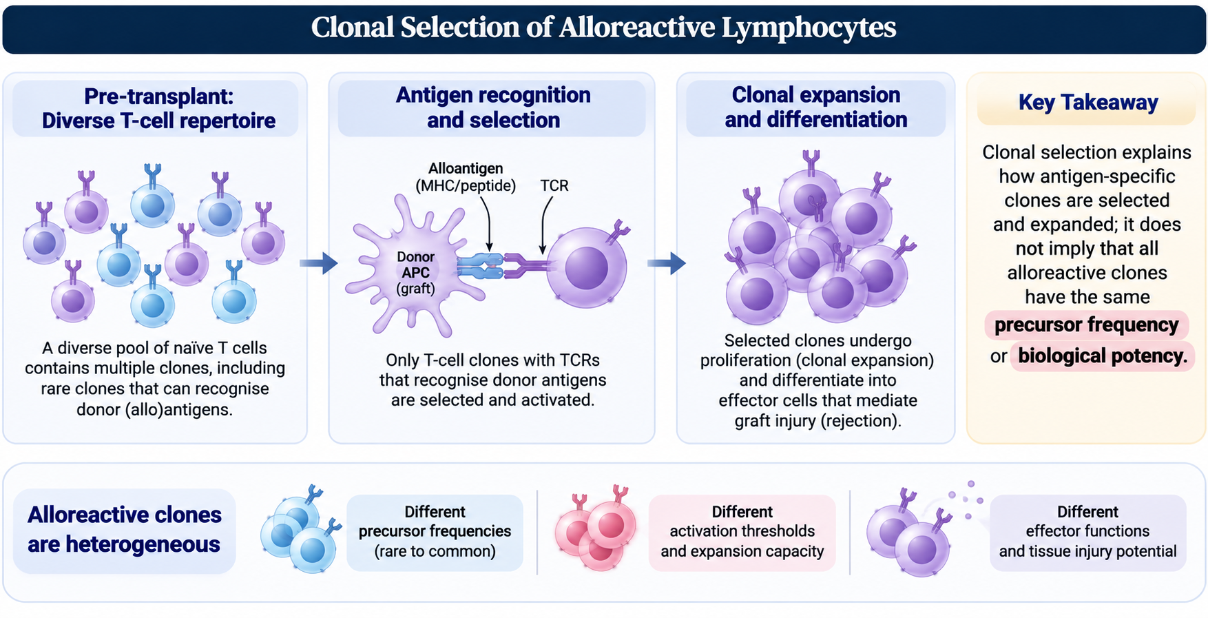
The genetic observations were later integrated with the clonal selection framework: lymphocytes bear antigen receptors with distinct specificities, and antigen exposure selectively expands the clones capable of recognising the relevant target. This provides a mechanistic explanation for both primary rejection and the accelerated second-set response. T cells may recognise intact donor MHC-peptide complexes directly or recognise donor-derived peptides presented by recipient MHC molecules; B cells can bind native donor antigens and differentiate into antibody-secreting plasma cells. The genetic basis of rejection therefore feeds directly into antigen recognition, clonal expansion and immune memory - the central mechanisms developed in Modules 1.2 and 1.3.
Burnet’s clonal-selection theory and transplantation
Burnet’s clonal-selection theory, formally articulated in 1957, proposed that lymphocytes carry distinct antigen specificities and that an antigen selectively activates and expands the clones able to recognise it. The theory supplied a cellular framework for understanding why a first graft primes a restricted population of donor-reactive lymphocytes and why a subsequent graft bearing the same alloantigens can trigger a faster response. In contemporary terms, alloreactive T-cell clones recognise intact donor HLA-peptide complexes or donor-derived peptides presented by recipient HLA, while B-cell clones can recognise native donor HLA or other graft antigens and generate antibodies. Burnet’s clonal-selection paper and the 1960 Nobel recognition of Burnet and Medawar for acquired immunological tolerance are related landmarks, but they are not the same discovery or Nobel citation.
1.1.4 Discovery of Histocompatibility and the Major Histocompatibility Complex
The murine H-2 system
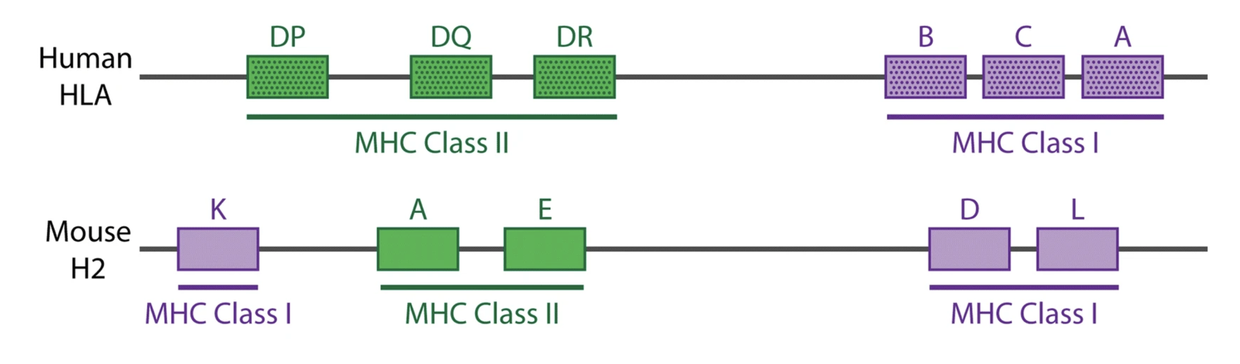
Gorer’s serological studies and Snell’s genetic analysis converged on the murine H-2 histocompatibility system. The H-2 region became the experimental prototype for the major histocompatibility complex (MHC). Inbred mouse strains and congenic strains - animals that differ at selected genetic regions while sharing much of the remainder of the genome - made it possible to identify loci with strong effects on graft rejection. The MHC earned its name because of its major influence on tissue and tumour compatibility, not because it is the only genetic determinant of transplantation outcomes.
From histocompatibility locus to immune-response system
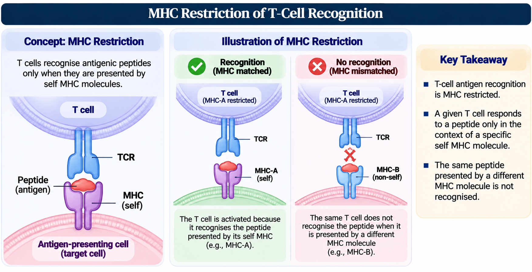
The MHC was initially identified through transplantation phenotypes, but subsequent experiments revealed its broader role in immune recognition. Baruj Benacerraf and colleagues identified immune-response genes linked to MHC in experimental animals. Later work showed that T cells recognise antigenic peptides in the context of MHC molecules, a principle termed MHC restriction. The Nobel Prize in Physiology or Medicine in 1980 recognised the contributions of Baruj Benacerraf, Jean Dausset and George Snell to discoveries concerning genetically determined cell-surface structures that regulate immune reactions. These findings transformed the MHC from a transplantation locus into a central organising system for adaptive immunity.
MHC restriction: Zinkernagel and Doherty
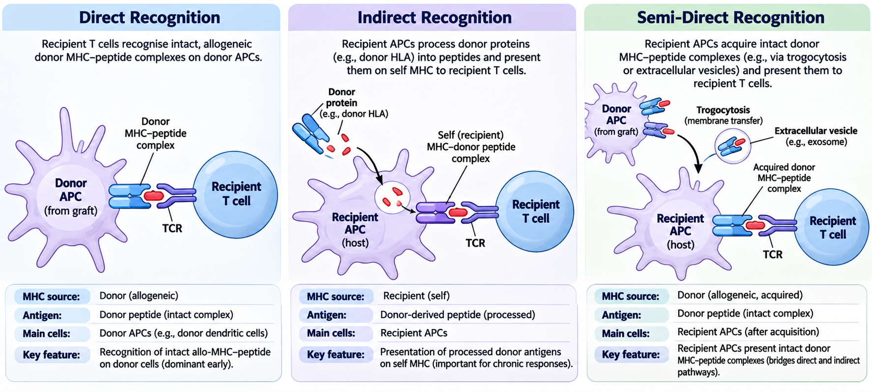
In 1974, Rolf Zinkernagel and Peter Doherty demonstrated that virus-specific cytotoxic T cells recognised infected target cells in the context of the appropriate self-MHC molecule. This principle, termed MHC restriction, showed that T-cell antigen recognition is not simply recognition of a foreign peptide in isolation: the T-cell receptor recognises a composite peptide-MHC surface. The discovery connected transplantation genetics to the fundamental mechanism of T-cell recognition and was recognised by the 1996 Nobel Prize in Physiology or Medicine. In transplantation, direct allorecognition is a special case in which recipient T cells can respond to intact allogeneic MHC-peptide complexes, while indirect allorecognition involves donor-derived peptides presented by recipient MHC.
Class I and class II: the conceptual framework
MHC class I molecules are expressed on most nucleated cells and present peptide antigens to CD8 T cells. MHC class II molecules are constitutively expressed by professional antigen-presenting cells and present peptides to CD4 T cells; their expression can be induced on other cell types under inflammatory conditions. In transplantation, class I and class II disparities can both provoke strong alloreactive responses. Direct recognition of intact donor MHC-peptide complexes can activate recipient T cells, while donor antigens processed by recipient antigen-presenting cells drive indirect recognition. These pathways ultimately connect the historical discovery of histocompatibility with contemporary mechanisms of T-cell-mediated rejection and DSA formation. Semi-direct antigen presentation occurs when recipient antigen-presenting cells acquire intact donor MHC–peptide complexes through mechanisms such as trogocytosis or extracellular vesicle (exosome)-mediated transfer, allowing them to present intact allogeneic MHC–peptide complexes to recipient T cells. This pathway combines features of direct and indirect allorecognition.
Why MHC polymorphism matters
MHC genes are highly polymorphic. Allelic variation changes the structure of the peptide-binding groove, the peptide repertoire that can be presented, the surface exposed to alloreactive T-cell receptors and the population distribution of HLA molecules. Consequently, a donor-recipient mismatch can be recognised through multiple routes: direct recognition of allogeneic MHC, recognition of donor peptides bound to self MHC, and antibody recognition of polymorphic HLA epitopes. This explains why the MHC is such a potent transplantation barrier and why HLA typing is central to donor selection, particularly in HCT.
Beyond the MHC
The discovery of the MHC did not eliminate the importance of other antigens. Minor histocompatibility antigens, ABO blood-group antigens, MICA and other non-HLA targets can contribute to incompatibility or graft injury. NK-cell receptors, including KIR, interact with HLA ligands and influence innate alloreactivity. MHC class III genes also encode immune mediators, including complement components and inflammatory proteins. Modern histocompatibility therefore considers HLA in a broader immunogenetic landscape rather than treating HLA matching as a complete measure of biological compatibility.
1.1.5 Discovery and Characterisation of the Human Leukocyte Antigen System
Human leukocyte antibodies revealed polymorphic antigens
The human MHC was identified through studies of antibodies against leukocytes. In the 1950s, investigators studying sera from multiply transfused patients and multiparous women observed antibody patterns that reacted with leukocytes from some individuals but not others. These findings showed that human leukocytes expressed inherited alloantigens. The immune sera were valuable reagents because pregnancy and transfusion had provided natural alloimmunisation events, effectively exposing HLA polymorphism before the molecular nature of the system was known.
Dausset, van Rood and Payne
Jean Dausset described the leukocyte antigen he called MAC in 1958, later recognised as HLA-A2. Dausset studied sera from multiply transfused patients and identified reproducible agglutination of leukocytes from selected donors. Jon van Rood and Rose Payne independently advanced the field using leukocyte antibodies, including sera from multiparous women, to define additional human leukocyte specificities and patterns of inheritance. Their work demonstrated that the antigen system was polymorphic and genetically controlled.
The mixed lymphocyte reaction and the HLA class II story
The mixed lymphocyte reaction (MLR; also called mixed lymphocyte culture, MLC) was independently described by Bach and colleagues and by Bain and colleagues in 1964. When leukocytes from genetically different individuals are cultured together, responder lymphocytes proliferate and undergo blast transformation. Family studies subsequently showed little or no response between HLA-identical siblings, linking the reaction to HLA disparity. The assay was an important functional tool for histocompatibility research and helped direct attention toward HLA-linked class II determinants, later characterised as HLA-DR, -DQ and -DP. MLC should be distinguished from a physical crossmatch: MLC measures cellular proliferative alloreactivity in vitro, whereas a crossmatch tests whether recipient serum reacts with donor cells. Neither is interchangeable with molecular HLA typing.
HLA genomic organisation, haplotypes and linkage disequilibrium
The human MHC is located on chromosome 6p21.3. The classical class I loci HLA-A, -B and -C lie in the class I region; the major class II loci include HLA-DR, -DQ and -DP; and the class III region contains genes encoding several complement components and inflammatory mediators rather than classical peptide-presenting HLA molecules. The extended MHC also includes non-classical HLA and MHC-associated genes, including MICA and MICB. HLA genes are highly polymorphic, and alleles at neighbouring loci are often inherited together because of physical linkage and population-specific linkage disequilibrium.
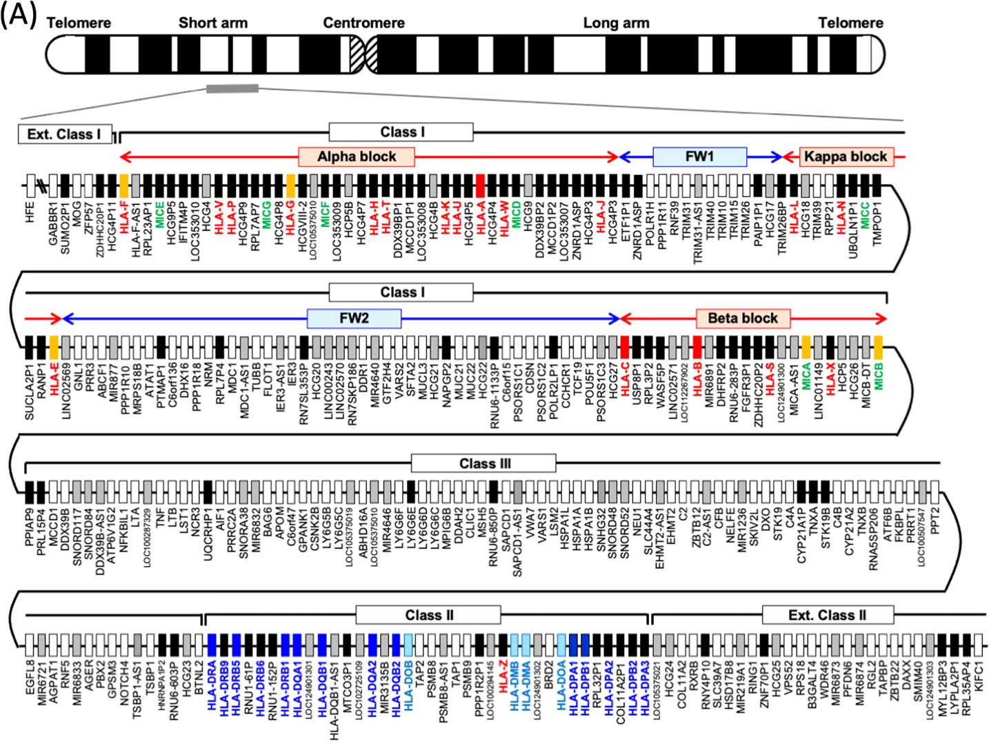
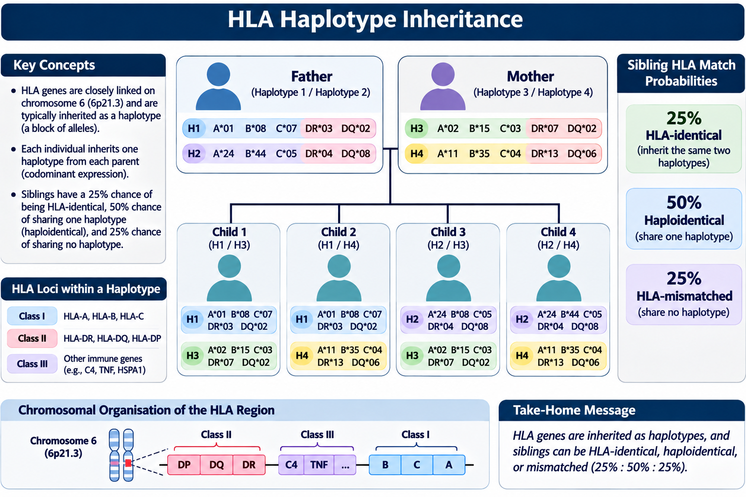
A haplotype is the linked set of HLA alleles inherited from one parent. HLA expression is codominant: the maternally and paternally inherited alleles are both expressed. In the simplified model without recombination, each parent transmits one of two haplotypes, so full siblings have a 25% probability of sharing both parental haplotypes, a 50% probability of sharing one and a 25% probability of sharing neither. Recombination can create exceptions, and linkage disequilibrium means that allele combinations occur at frequencies that vary among populations. These principles underpin family studies, haplotype inference, related-donor selection and population-aware interpretation of HLA match probabilities.
HLA nomenclature and sequence reference resources
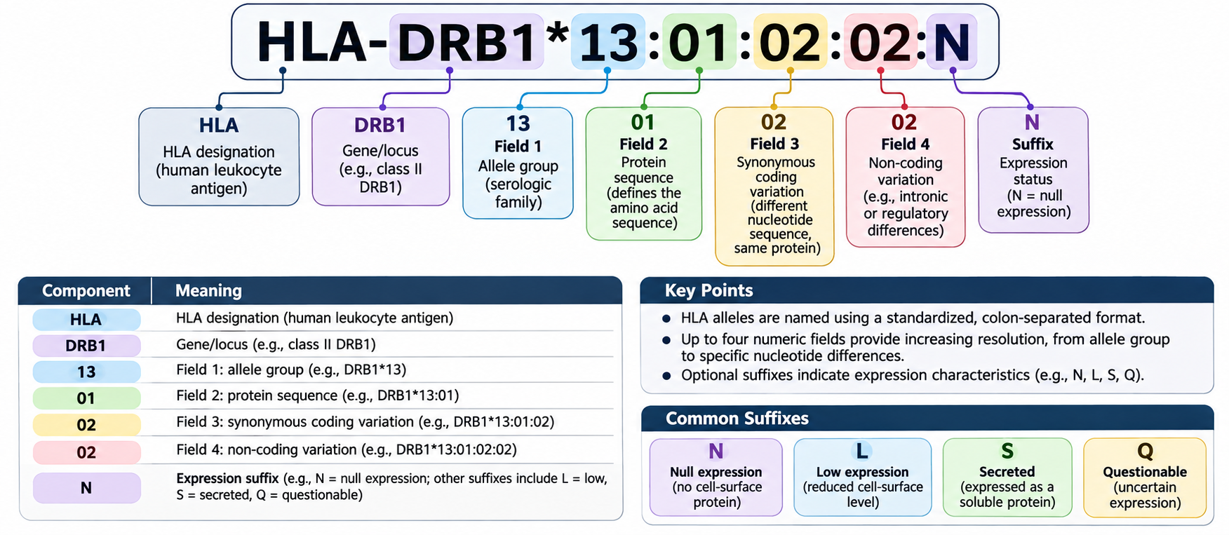
As serological specificities gave way to DNA-defined alleles, internationally coordinated nomenclature became essential. The WHO Nomenclature Committee for Factors of the HLA System has maintained standardised gene and allele designations since the late 1960s. The IPD-IMGT/HLA Database, first released in 1998, curates HLA allele sequences and supports consistent allele identification across laboratories and research groups. At the time of this revision, the database lists Release 3.66 (October 2026); allele assignment and reporting should always identify the relevant database release and typing resolution when these details affect interpretation. A gene-level or allele-level label, serological antigen assignment and expression suffix are not interchangeable descriptions.
International workshops, nomenclature and shared reagents
The growing complexity of HLA specificities required international coordination. Histocompatibility workshops allowed laboratories to compare sera, cells, typing methods and antigen assignments. They supported the development of shared nomenclature, reference reagents, family studies, donor registries and databases. As the number of recognised alleles expanded and molecular typing became possible, the nomenclature had to evolve from serological antigen assignments to allele-level sequence definitions. The current HLA system is thus both a biological discovery and an international standardization achievement.
From serological antigens to molecular alleles
Early HLA typing depended on antibodies that recognised expressed cell-surface antigens. This approach was foundational but constrained by limited reagent specificity, shared epitopes, weak or null expression, population bias in available antisera and the difficulty of distinguishing closely related alleles. DNA-based typing later enabled direct characterisation of HLA gene polymorphism. Contemporary high-resolution and next-generation sequencing methods can resolve differences that serological typing could not identify, including protein-level and synonymous or noncoding variation when the assay covers the relevant regions. Oberle’s modern guide and Eisenbrey’s historical account show how laboratory practice shifted from cell-based serology toward molecular typing while retaining the clinical relevance of expressed antigen and antibody reactivity.
Clinical significance of HLA
HLA molecules are central to graft compatibility because they are highly polymorphic and are expressed on donor cells, including graft endothelium and antigen-presenting cells. In solid-organ transplantation, HLA mismatch is one component of immunological risk and its impact is modified by organ type, urgency, donor availability, DSA and immunosuppressive therapy. In HCT, HLA compatibility is especially important because donor-derived immune cells must engraft in a recipient whose tissues may express different HLA molecules. The discovery of HLA therefore enabled the development of tissue typing, donor selection, antibody screening and crossmatching, while also opening the way to studying HLA-restricted immune responses, disease associations and pharmacogenetics. [8, 10, 12, 13, 18]
1.1.6 Discovery of Acquired Immunological Tolerance
Natural chimerism in cattle twins
A key clue to acquired tolerance came from Raymond Owen’s 1945 study of dizygotic cattle twins. Through shared placental vascular connections, twins could exchange haematopoietic cells and develop persistent blood-cell chimerism. Despite carrying genetically distinct blood-cell populations, the twins could tolerate one another’s cells. This natural biological experiment suggested that exposure to foreign cells early in development could alter the later immune response to those cells. It provided a foundation for experimental work on acquired immunological tolerance. Later, Billingham, Brent and Medawar tested whether early exposure to foreign cells could induce specific acceptance of donor tissues. In their landmark 1953 Nature paper, they showed that neonatal exposure to foreign cells could produce a state in which the recipient later accepted grafts from the same donor strain. The tolerance was acquired, antigen-specific and dependent on the developmental context of exposure. The result provided experimental support for the concept that immune unresponsiveness to selected antigens could be induced rather than being solely an inherited or passive state.
From acquired tolerance to central and peripheral tolerance
The original experiments preceded modern knowledge of lymphocyte development and regulatory networks. Subsequent work established that central tolerance is shaped during T-cell development in the thymus and B-cell development in the bone marrow, while peripheral tolerance controls potentially autoreactive or alloreactive lymphocytes that escape developmental selection. Mechanisms include deletion, anergy, suppression by regulatory T cells, inhibitory receptor signalling and specialised tissue environments. Janeway emphasizes the multiple layers of tolerance that normally prevent self-reactivity and the possibility that their failure produces disease; the same conceptual framework helps explain why graft-specific tolerance is biologically plausible but difficult to achieve.
Tolerance is not equivalent to immunosuppression
Pharmacological immunosuppression broadly reduces immune activation or effector function and generally requires ongoing treatment to prevent rejection. Operational tolerance describes stable graft function in the absence of maintenance immunosuppression, without evidence of active rejection, in carefully defined clinical settings. Donor-specific tolerance is a more restrictive concept: immune reactivity is selectively restrained toward donor antigens while protective immunity against unrelated antigens is preserved. The distinction matters because drug-induced suppression may control rejection without reprogramming the underlying antigen-specific immune repertoire. Clinical tolerance strategies seek to alter that balance rather than simply intensify global immune suppression.
Chimerism as a translational strategy
The tolerance work informed strategies that combine donor haematopoietic cells with recipient immunity to establish mixed or full donor chimerism. Donor-derived antigen-presenting cells can participate in central deletion of donor-reactive lymphocytes, while peripheral regulatory mechanisms can maintain unresponsiveness. In some experimental and highly selected clinical protocols, combined haematopoietic cell and solid-organ transplantation has been used to pursue donor-specific tolerance. These approaches remain specialised and carry risks, including conditioning toxicity, graft failure and GVHD. They are not equivalent to routine withdrawal of immunosuppression.
The fetus as a naturally tolerated semi-allogeneic conceptus
Medawar used the maternal-fetal relationship to frame a central immunological question: why is a genetically distinct, semi-allogeneic fetus not ordinarily rejected by the maternal immune system? The concept is often called the fetal allograft paradox. Subsequent work has shown that pregnancy is supported by specialised regulation at the maternal-fetal interface, including trophoblast biology, local innate and adaptive immune regulation, vascular remodelling and temporally coordinated inflammatory processes. This is not equivalent to global maternal immunosuppression, nor is the fetus simply an organ graft. The comparison is useful because it illustrates that antigenic disparity alone does not determine outcome; tissue context and local regulation are decisive.
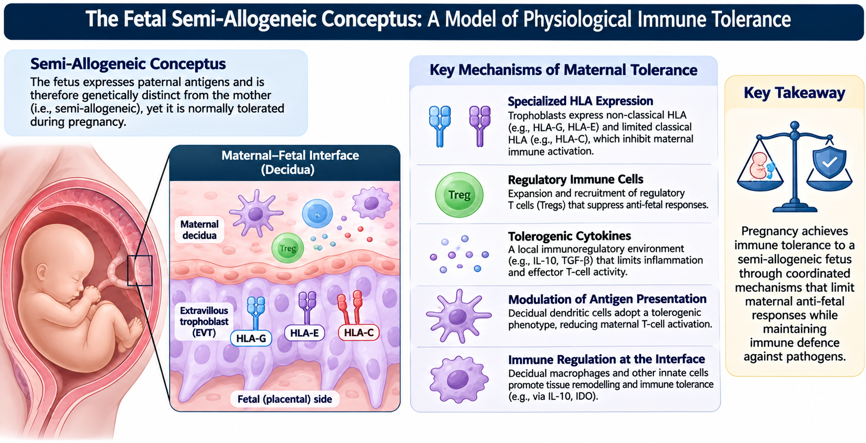
1.1.7 Development of Experimental Organ Transplantation
Why experimental organ models were necessary
Skin grafts established the principles of rejection, but transplantation of vascularised organs introduced additional biological variables: vascular endothelium, parenchymal cell types, organ-specific antigen expression, ischaemia-reperfusion injury, innate immune activation and the consequences of organ dysfunction. Experimental organ transplantation allowed investigators to study vascularised grafts in controlled settings and to separate technical failure from immune-mediated injury. It also enabled testing of immunosuppressive interventions before clinical application. Li and Jevnikar emphasise the continuing importance of experimental models for discovery and translation, while warning that model-specific findings cannot be assumed to predict human outcomes directly.
Vascularised organ transplantation in animals
Advances in vascular anastomosis enabled kidney, heart and other organ transplantation in experimental animals. Heterotopic models place the graft in a non-native anatomical position while retaining its vascular supply; orthotopic models replace the native organ in its usual anatomical position. For example, heterotopic heart transplantation can provide a technically accessible graft for serial assessment, whereas orthotopic models better reproduce the physiological role of the organ. Kidney, liver, lung and heart models each provide different insights because the grafts differ in vascular anatomy, parenchymal vulnerability, antigen-presenting cell populations and functional readouts.
Experimental variables and model interpretation
A transplant model is defined by more than the organ. Important variables include species and strain, donor-recipient genetic disparity, graft site, cold and warm ischaemia, preservation method, recipient conditioning, immunosuppressive regimen, microbiological status and the timing of tissue sampling. A strong allogeneic barrier in one strain combination may be much weaker in another. Graft survival alone is an incomplete endpoint: histopathology, immune-cell infiltration, donor-specific antibodies, complement activation, graft function and molecular signatures provide complementary evidence. These considerations are essential when interpreting experimental results and when deciding whether a model addresses acute rejection, chronic injury, tolerance or a specific effector pathway.
The value and limits of animal models
Small-animal models provide genetic control, access to defined immune pathways and the ability to perform mechanistic interventions. Their limitations include strain-specific responses, differences in immune-cell composition and organ physiology, and the compressed timescale of some experimental diseases. Large-animal models can improve anatomical and physiological translation and may be valuable for surgical innovation, device testing and selected immunological questions, but are more resource-intensive. Humanised models and ex vivo systems add further approaches, although they do not fully recreate the human immune system or the tissue microenvironment. Experimental findings should therefore be interpreted as evidence within a defined model, not as direct proof of clinical efficacy.
From experimental transplantation to translational questions
Experimental organ models helped define the roles of T cells, B cells, macrophages, dendritic cells, antibodies, complement and endothelium in graft injury. They also facilitated the development of immunosuppressive drugs and tolerance strategies. Contemporary experiments extend beyond survival to tissue-resolved and molecular readouts, including transcriptomics, single-cell profiling, spatial analysis and donor-derived biomarkers. The historical purpose of experimental transplantation remains unchanged: to establish causal mechanisms under controlled conditions, then test whether those mechanisms are relevant in human transplantation.
1.1.8 Evolution of Clinical Organ Transplantation and Immunosuppression
The transition from operative success to durable clinical therapy
Clinical organ transplantation required convergence of surgical technique, organ preservation, donor selection, blood-group compatibility, infection control and management of alloimmunity. The kidney became the first widely established solid-organ transplant because it could be surgically implanted, its function could be assessed biochemically, and dialysis offered a bridge or alternative in selected patients. The 1954 Boston kidney transplant, performed by Joseph E. Murray with John P. Merrill, J. Hartwell Harrison and their clinical team between identical twins, is widely recognised as the first durable, documented human kidney transplant, demonstrating that a vascularised organ could function long term when major genetic disparity was absent. A Canadian historical review also describes Gordon Murray’s 1952 deceased-donor kidney procedure in Toronto, with long reported recipient survival, but notes that documentation and follow-up limitations have left the claim debated. This is a useful example of why a historical “first” requires defined evidentiary criteria.
Early allogeneic transplantation and immunosuppression
The next challenge was transplantation between genetically non-identical individuals. Early strategies included total-body irradiation and other forms of lymphoid depletion, but toxicity was substantial. Antimetabolites such as 6-mercaptopurine and azathioprine reduced lymphocyte proliferation; corticosteroids provided broad anti-inflammatory and immunosuppressive effects. Combination regimens improved the ability to control acute rejection. Polyclonal antilymphocyte and antithymocyte preparations were subsequently used to deplete or suppress T cells, while monoclonal antibodies offered more selective cellular targets. These advances changed transplantation from a test of surgical feasibility into a discipline in which immune responses could be manipulated pharmacologically.
Historical progression of pharmacological immunosuppression
The pharmacological history is best understood as a sequence of increasingly selective ways to interrupt lymphocyte activation and expansion. Early clinical efforts included irradiation, corticosteroids and antimetabolites, but the therapeutic window was narrow. 6-mercaptopurine and azathioprine were developed by Gertrude Elion and George Hitchings; Roy Calne, Joseph Murray and other investigators translated these compounds into experimental and clinical transplantation. Early human attempts with 6-mercaptopurine were not uniformly successful, and durable progress followed combination approaches that paired azathioprine with corticosteroids.
Cyclosporine and the calcineurin-inhibitor era
Borel and colleagues reported the immunosuppressive effects of cyclosporine A in 1976. Calne and colleagues then reported its early clinical use in recipients of cadaveric renal allografts in 1978. These developments transformed solid-organ transplantation. Cyclosporine binds cyclophilin; the drug-immunophilin complex inhibits calcineurin, thereby reducing NFAT activation and transcription of IL-2 and other T-cell activation genes. Tacrolimus (FK506) binds FKBP12 and also inhibits calcineurin, providing a related but pharmacologically distinct approach. Calcineurin inhibitors became central components of maintenance immunosuppression and contributed to improved graft survival and expansion of transplantation to additional organs. Their use also highlighted the trade-offs of chronic immunosuppression, including nephrotoxicity, infection, malignancy and metabolic complications.
Expansion of the immunosuppressive armamentarium
Mycophenolate mofetil inhibits inosine monophosphate dehydrogenase and limits de novo guanine nucleotide synthesis, particularly affecting proliferating lymphocytes. Sirolimus and everolimus bind FKBP12 but inhibit mTOR, limiting growth-factor-driven cell-cycle progression. Induction strategies may include lymphocyte-depleting agents or antibodies targeting selected immune pathways. Modern regimens are combinations chosen according to organ type, immunological risk, recipient comorbidity, toxicity profile and the phase after transplantation. These drugs are not mechanistically interchangeable: they act at different stages of lymphocyte activation, proliferation and effector function.
Organ-specific clinical milestones and programme development
Clinical transplantation expanded beyond the kidney. Early liver transplantation attempts were developed by Thomas Starzl and colleagues in the 1960s, with more durable success emerging as surgical technique and immunosuppression improved. Christiaan Barnard performed the first human heart transplant in 1967. Lung transplantation faced additional challenges related to airway healing, infection and chronic graft dysfunction, with reliable long-term outcomes developing later. Pancreas, intestine and composite tissue transplantation broadened the clinical scope. Each organ presents distinct immunological and non-immunological challenges, so success cannot be attributed to immunosuppression alone.
The persistent balance between rejection and over-immunosuppression
The clinical objective is not maximal immune suppression. It is sufficient control of alloimmunity to preserve graft function while retaining protective responses against infection and malignancy. T-cell-mediated rejection, antibody-mediated rejection and chronic graft injury differ in pathogenesis and response to therapy. Conventional immunosuppression is more effective against some T-cell pathways than against established antibody-mediated injury or chronic microvascular damage. Consequently, modern care increasingly combines donor selection, antibody assessment, biopsy interpretation, immune monitoring and tailored immunosuppression. The historical progress from broad suppression toward mechanism-specific management is a central theme of precision transplantation.
1.1.9 Development of Bone Marrow and Haematopoietic Cell Transplantation
Radioprotection and marrow rescue
The foundations of HCT arose from experiments investigating recovery after lethal irradiation. Jacobson and colleagues showed that shielding the spleen could protect animals from otherwise lethal radiation exposure, implicating a radiosensitive haematopoietic compartment. Lorenz and colleagues then demonstrated that infusion of bone marrow cells could protect irradiated mice and guinea pigs. These experiments established that marrow contained cells capable of restoring haematopoiesis. Later lineage-tracing and transplantation studies clarified that durable recovery depended on donor-derived haematopoietic stem and progenitor cells rather than simply transient support by mature blood cells.
Till and McCulloch: quantitative evidence for clonogenic marrow cells
Till and McCulloch’s 1961 work developed a direct assay of radiation sensitivity by quantifying colony-forming units in the spleen (CFU-S) after transplantation of normal mouse marrow into irradiated recipients. The appearance of discrete colonies provided evidence that individual marrow-derived clonogenic progenitors could proliferate and generate haematopoietic descendants. This was a decisive foundation for stem-cell biology, but the assay should not be described as a complete modern prospective definition of long-term, self-renewing haematopoietic stem cells; later lineage, serial-transplantation and phenotypic studies refined that concept.
Early clinical marrow infusion
E. Donnall Thomas and colleagues pursued marrow infusion in patients with severe haematological disease during the 1950s. Early attempts demonstrated that donor marrow could sometimes engraft after conditioning, but outcomes were limited by graft failure, infection, organ toxicity and severe GVHD. Thomas’s 1957 clinical report is an important milestone in translating marrow rescue to humans, but it should not be equated with the establishment of durable allogeneic HCT as a routine therapy. Clinical success required better donor selection, conditioning regimens, supportive care, infection control and understanding of donor-host immunobiology.
Canine models and the move to successful allogeneic HCT
The canine model was central to the transition from marrow rescue experiments to reproducible allogeneic HCT. Thomas and colleagues demonstrated marrow engraftment in dogs in the early 1960s, while Storb and colleagues used defined dog leukocyte antigen (DLA)-matched and mismatched donor-recipient pairs to study engraftment, rejection, GVHD and the effects of conditioning. The large-animal model allowed systematic evaluation of donor selection and immunoprophylaxis and helped shape principles later translated to human HCT.
1968: allogeneic marrow transplantation for SCID
Gatti and colleagues reported immunological reconstitution after allogeneic bone marrow transplantation in an infant with sex-linked severe combined immunodeficiency in 1968. This is widely recognised as a first successful allogeneic HCT milestone for SCID, although early cases also exposed the risk of GVHD and the importance of donor compatibility. The landmark should be distinguished from Thomas’s earlier clinical marrow infusions in the 1950s, which were important translational attempts but did not yet establish durable allogeneic HCT as a reliable therapy.
HCT as replacement and immune reconstitution
Unlike solid-organ transplantation, HCT introduces donor haematopoietic stem cells and a variable inoculum of mature donor immune cells into a conditioned recipient. The intended outcome is donor-derived haematopoiesis and immune reconstitution. HCT can treat haematological malignancies, bone-marrow failure syndromes, selected inherited blood disorders, immunodeficiencies and metabolic diseases. Conditioning chemotherapy and/or irradiation reduces malignant or host haematopoietic cells, suppresses host immunity and creates conditions that permit donor-cell engraftment. The balance between disease control, toxicity, engraftment and immune complications differs across myeloablative, reduced-intensity and non-myeloablative approaches. [9, 10]
HLA matching, donor sources and access
Recognition that HLA disparity strongly influences alloreactivity transformed donor selection. HLA-identical sibling transplantation became an important route to clinical success, followed by the development of unrelated donor registries and cord-blood banks. Bone marrow was the original graft source; mobilised peripheral-blood stem cells later became widely used and often permit faster haematopoietic recovery, although graft composition and chronic GVHD risk can differ. Umbilical cord blood broadened donor access and can tolerate some HLA disparity, but cell dose and delayed immune recovery create specific constraints. Haploidentical related donors and post-transplant cyclophosphamide expanded donor options for patients without a matched sibling or unrelated donor.
Cord blood and unrelated donor registries
The first successful umbilical-cord-blood transplant was performed in 1988 in a child with Fanconi anaemia using HLA-identical sibling cord blood; the clinical report appeared in 1989. Cord blood broadened access to allogeneic haematopoietic cells and can permit some HLA disparity, although cell dose and delayed immune recovery remain important constraints. Unrelated-donor transplantation also expanded through national and international registries. NMDP’s historical account describes the first unrelated-donor marrow transplant in 1979 and its first registry-coordinated transplant in December 1987; donor registries subsequently became a major route to unrelated volunteer grafts.
HCT and solid-organ transplantation are immunologically distinct
In solid-organ transplantation, the principal immune challenge is the recipient response against donor tissue. In allogeneic HCT, donor immune cells can attack recipient tissues, and host immune cells can reject the donor graft. HCT therefore includes bidirectional alloreactivity: graft rejection or graft failure, GVHD, and beneficial GVL activity. This reciprocal structure explains why HLA matching and T-cell control are especially important in HCT and why graft source, T-cell content, conditioning and prophylaxis are integral to transplant design.
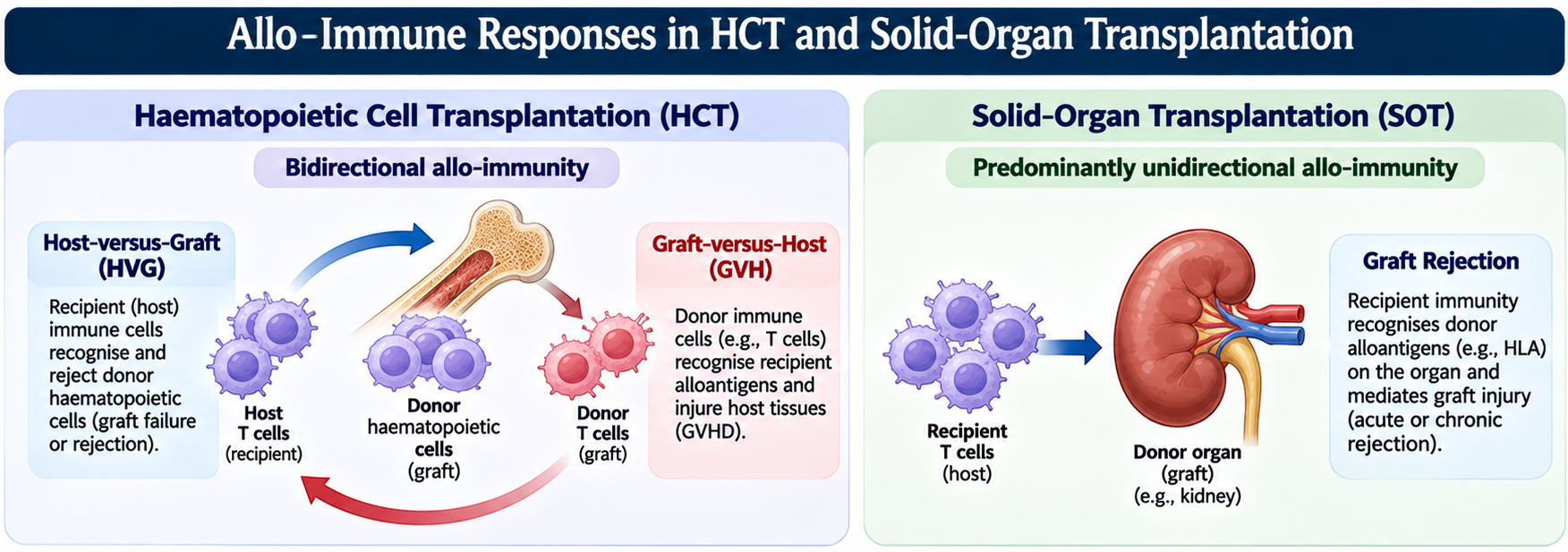
1.1.10 Discovery of Graft-versus-Host Disease and Graft-versus-Leukaemia Effects
The biological logic of GVHD
GVHD occurs when immunocompetent donor lymphocytes recognise recipient antigens as foreign and attack host tissues. Classic experimental criteria include the transfer of immunologically competent cells, antigenic disparity between donor and recipient, and insufficient host capacity to eliminate the donor immune cells. The syndrome became apparent during early marrow transplantation attempts and was clarified by animal experiments involving irradiation and donor marrow. Barnes and Loutit’s studies helped demonstrate that donor-derived immune cells could produce a systemic “secondary disease” after marrow transplantation. Simonsen and other investigators contributed to the experimental analysis of this phenomenon.
Clinical observation and mechanistic confirmation
Early clinical HCT reports described severe post-transplant illness characterised by skin, gastrointestinal and hepatic injury. In a 1965 report by Mathé and colleagues, donor marrow engraftment was associated with a syndrome later understood as GVHD, while leukaemia remained in remission for a period. These observations helped reveal that donor alloreactivity could be both harmful and therapeutically beneficial. Modern understanding recognises that GVHD risk depends on HLA disparity, minor histocompatibility antigens, donor T-cell content, conditioning-related tissue injury, inflammatory signals, microbiome disruption and recipient factors.
Tissue targets and effector mechanisms
Acute GVHD commonly affects skin, gastrointestinal tract and liver. Donor CD4 and CD8 T cells become activated in secondary lymphoid tissues through interactions with recipient antigen-presenting cells and then traffic to target organs. Tissue damage is mediated through cytotoxic pathways, inflammatory cytokines and recruitment of innate effector cells. Conditioning-related injury can amplify the inflammatory environment by releasing damage-associated signals and increasing antigen presentation. Chronic GVHD is biologically more heterogeneous and may include immune dysregulation, fibrosis, autoantibody-like processes and tissue remodelling. The clinical phenotype therefore cannot be reduced to a single pathway.
The graft-versus-leukaemia effect
GVL describes donor immune activity against residual malignant haematopoietic cells after HCT. Donor T cells and NK cells can recognise tumour-associated antigens, minor histocompatibility antigens selectively expressed by haematopoietic cells, or other tumour-associated targets. This effect contributes to the curative potential of allogeneic HCT in leukaemia and some other malignancies. The central therapeutic problem is to preserve clinically meaningful GVL activity while preventing or limiting GVHD. The two effects are related but not identical: they can share effector populations and antigenic targets, yet their tissue distribution, antigen specificity and susceptibility to immune regulation may differ.
Strategies to separate beneficial and harmful alloreactivity
HCT prophylaxis and graft engineering attempt to reduce GVHD without abolishing GVL. Approaches include calcineurin inhibitors combined with methotrexate or mycophenolate, antithymocyte globulin, ex vivo T-cell depletion, selective depletion of subsets, regulatory-cell strategies and post-transplant cyclophosphamide. These interventions differ in their effects on alloreactive T cells, immune reconstitution, infection risk and relapse. Post-transplant cyclophosphamide has particularly broadened haploidentical transplantation by selectively reducing the expansion of activated alloreactive lymphocytes while permitting subsequent immune recovery. No approach entirely eliminates the trade-off between GVHD, infection, relapse and graft failure.
1.1.11 Development of Modern Histocompatibility Testing and HLA Matching
The serological era
The first clinical HLA methods relied on antibodies that recognised cell-surface antigens. Leukoagglutination studies helped identify human leukocyte alloantigens, and the microlymphocytotoxicity assay developed by Terasaki and McClelland in 1964 provided a practical standard for detecting HLA antigens and antibodies using viable lymphocytes, patient or reference serum and complement. Cell lysis was measured with vital dyes. The same underlying assay principle supported serological typing, antibody screening and physical crossmatching. It was a major advance because it converted qualitative observations into a standardised laboratory method that could be shared between centres.
Crossmatching and prevention of hyperacute rejection
A physical crossmatch tests whether recipient serum contains antibodies that bind donor lymphocytes. In a complement-dependent cytotoxicity crossmatch, donor cells are incubated with recipient serum and complement; antibody-mediated cell lysis indicates potentially dangerous preformed reactivity. Flow cytometric crossmatching later increased sensitivity by detecting cell-bound immunoglobulin even when complement-mediated lysis was not observed. The pretransplant crossmatch became a critical safety procedure because preformed donor-reactive antibodies can bind vascular endothelium and cause rapid graft injury, including hyperacute rejection. ABO compatibility and careful donor-recipient identification remain separate but essential safeguards.
Patel and Terasaki: the clinical significance of a positive crossmatch
In 1969, Patel and Terasaki reported that a positive pretransplant lymphocyte crossmatch was strongly associated with immediate kidney-graft failure. In their landmark series, immediate failure occurred in 24 of 30 recipients with a positive crossmatch compared with 8 of 195 recipients with a negative result. The study made clear that circulating preformed donor-reactive antibodies could cause catastrophic early graft injury and established the crossmatch as a major transplant-safety procedure. The assay’s predictive meaning depends on the cell target, immunoglobulin class, complement activity, assay sensitivity, treatment history and clinical context; a positive result must be interpreted rather than treated as a context-free binary.
From cellular to molecular HLA typing
Serological typing was limited by reagent availability, shared epitopes, variable antigen expression, difficulty typing class II antigens and the inability to resolve allelic differences invisible to antibodies. Molecular methods changed this landscape. Early DNA-based approaches included restriction fragment length polymorphism; PCR enabled sequence-specific primer and sequence-specific oligonucleotide methods; sequencing-based typing provided direct allele sequence information; and next-generation sequencing enabled simultaneous high-resolution analysis of multiple HLA loci. Long-read sequencing and phasing approaches can improve resolution of haplotypes and complex ambiguities. Oberle’s guide details the current methodological spectrum, including short-read and long-read sequencing, while Eisenbrey traces the transition from serology to molecular typing.
HLA matching in solid-organ transplantation and HCT
HLA matching has different operational importance across transplant settings. In kidney transplantation, HLA compatibility can reduce alloimmune risk, but organ availability, urgency, living donation, antibody status, age, comorbidity and other clinical factors also determine allocation and acceptance. Modern immunosuppression has made transplantation possible across substantial HLA disparity. In HCT, HLA matching remains a major determinant of graft failure, GVHD and survival because donor immune cells directly encounter recipient tissues. The 2026 EBMT Practice Harmonisation and Guidelines Committee recommendations favour next-generation sequencing-based approaches and recommend at least high-resolution typing for HLA-A, -B, -C, -DRB1, -DQB1 and -DPB1, with additional loci and ultra-high-resolution strategies considered according to context, particularly when DSA may be relevant.
Antibody testing and the virtual crossmatch
The microcytotoxicity assay was followed by flow cytometric methods and then solid-phase assays using purified or recombinant HLA antigens attached to microbeads. Single-antigen bead assays can identify antibody reactivity at a much finer specificity level than cell panels, although mean fluorescence intensity is semiquantitative and assay artefacts require careful interpretation. The virtual crossmatch compares the recipient’s antibody profile with the donor’s HLA type to estimate compatibility without physically mixing donor cells and recipient serum. It complements rather than universally replaces physical crossmatching. Modern histocompatibility assessment therefore combines genotype, antibody specificity, assay limitations, sensitisation history, donor characteristics and clinical context.
From antigen-level matching to epitope and eplet analysis
Traditional antigen matching compares serologically defined HLA antigens; molecular typing resolves alleles and, depending on assay coverage, sequence-level differences. Epitope analysis asks whether donor HLA presents surface configurations that may be recognised by recipient antibodies. Eplets are algorithmically defined small configurations of polymorphic amino acids on the HLA molecular surface that contribute to antibody-accessible epitopes. Tools such as HLAMatchmaker can estimate eplet mismatches and support immunological risk assessment, particularly in kidney transplantation. However, a predicted eplet mismatch is not synonymous with a proven antibody response, and an eplet mismatch count is not a universal, validated substitute for HLA typing, DSA assessment or physical/virtual crossmatching. Clinical thresholds and predictive utility depend on the transplant setting, the algorithm and the evidence base.
Non-HLA antibodies: important targets, evolving evidence
Antibody-mediated graft injury is not restricted to HLA. Reported non-HLA targets include MICA/MICB, the angiotensin II type 1 receptor (AT1R), endothelin receptor A (ETAR) and other endothelial or tissue-associated antigens. Lymphocyte-based crossmatches do not detect every antibody directed against antigens that are not expressed on donor lymphocytes, including MICA in many assay formats. The clinical evidence, assay standardisation and treatment implications vary substantially across targets. A positive non-HLA antibody result alone should not establish antibody-mediated rejection or independently dictate therapy; interpretation requires integration with graft function, histopathology, HLA DSA, crossmatch findings and alternative causes of injury.
1.1.12 Modern Transplantation Immunology: From Rejection to Precision Transplantation
Beyond a binary model of rejection
Modern transplantation immunology no longer treats graft outcome as a binary choice between acceptance and rejection. Grafts experience a continuum of immune and non-immune processes, including ischaemia-reperfusion injury, infection, drug toxicity, recurrent disease, cellular rejection, antibody-mediated rejection, mixed phenotypes, chronic microvascular injury and fibrosis. A detectable immune response does not automatically establish tissue injury, and graft injury does not automatically identify its cause. Accurate interpretation requires integration of clinical function, histopathology, HLA antibody data, molecular evidence and temporal context.
Standardised pathology and clinicopathological integration
The Banff classification emerged from the first international consensus meeting for renal allograft pathology in 1991. Its purpose was to standardise biopsy interpretation, define reproducible lesion scores and create shared diagnostic categories for clinical practice and research. The current Banff Foundation reference guide for renal transplant pathology is version 2024-3, updated 30 September 2026, and incorporates iterative evidence-informed changes. Banff illustrates the movement from descriptive histology toward structured, integrated diagnosis, including histological lesions, serological findings and additional diagnostic parameters. It is a classification framework, not a substitute for clinical judgement or the assessment of alternative causes of graft dysfunction.
Molecular monitoring and biomarker interpretation
Transplant biomarker research has expanded from conventional serum creatinine and proteinuria toward donor-derived cell-free DNA, gene-expression signatures, urinary chemokines, donor-specific antibody characteristics and tissue transcriptomics. Donor-derived cell-free DNA can provide a non-invasive signal of graft-derived injury, but it is not specific for rejection: infection, ischaemia-reperfusion injury, drug toxicity and other forms of tissue damage can also increase levels. Fractional values can be influenced by recipient-derived cell-free DNA and clinical context. A biomarker therefore needs analytical validation, defined thresholds, prospective clinical validation and evidence that its use improves decisions or outcomes before it can replace biopsy or guide therapy on its own.
Systems immunology and tissue-resolved approaches
Single-cell RNA sequencing, CITE-seq, spatial transcriptomics, high-dimensional cytometry and computational integration enable analysis of immune-cell states and tissue compartments that were difficult to resolve with bulk assays. These approaches can distinguish infiltrating lymphocytes, resident immune cells, endothelial activation, stromal remodelling and local antigen-presentation programmes. Longitudinal profiling may reveal transitions from acute inflammation to chronic injury or operational tolerance. The principal challenge is translating molecular states into reproducible, clinically actionable classifications. High-dimensional data do not automatically solve causal inference, and a molecular signature associated with rejection is not necessarily a validated treatment-selection test.
Precision immunosuppression and tolerance-focused approaches
Precision transplantation seeks to match the intervention to the mechanism and risk profile. Examples include using HLA antibody and epitope information to define immunological risk; tailoring immunosuppression to rejection phenotype; monitoring drug exposure and immune status; and developing strategies that selectively regulate donor-reactive T cells, B cells or antibody-producing plasma cells. Tolerance-focused approaches include mixed chimerism, regulatory T-cell therapy, costimulation blockade and cellular engineering. These strategies remain at different levels of clinical maturity. A mechanistic rationale or promising early trial is not equivalent to established standard of care; evidence must be appraised by study design, reproducibility, safety and patient-important outcomes.
Xenotransplantation and engineered grafts
Xenotransplantation has returned to clinical attention through gene-edited pig organs designed to reduce major immunological incompatibilities and improve physiological compatibility. Recent clinical experiences with pig hearts and kidneys in selected living patients demonstrate that the field has progressed beyond preclinical models. However, the 2026 review by Cooper and colleagues notes that long-term patient and graft survival remain unresolved, and that thrombotic microangiopathy and proteinuria remain important concerns in renal xenotransplantation despite use of gene editing and costimulation blockade. Xenotransplantation is therefore an emerging translational field, not a routine alternative to human allografting.
The precision-transplantation paradigm
The modern goal is to understand the graft as an evolving biological system shaped by donor genetics, recipient immunity, tissue-resident and circulating immune cells, endothelial and stromal responses, treatment exposure, infection, microbiome and time after transplantation. The key questions are mechanistic and longitudinal: which target is recognised, by which immune component, in which anatomical compartment, with what evidence of injury, and what intervention is likely to change the outcome? This framework connects the historical discovery of rejection to contemporary immunogenetics, pathology, multi-omics and cell-based therapies. It also provides the conceptual bridge to the remaining modules of Pillar 1.
Integrative synthesis: what changed across the history?
The history of transplantation immunology can be understood as a sequence of increasingly precise explanations for graft outcome. First, vascular surgery made organ transfer technically feasible. Second, skin grafts provided an accessible model in which rejection could be observed and compared with autograft survival. Third, genetic studies identified inherited histocompatibility determinants. Fourth, H-2 and HLA research connected those determinants to antigen presentation and adaptive immune recognition. Fifth, tolerance experiments showed that the immune response could be altered in an antigen-specific manner. Finally, clinical immunosuppression, HLA typing, crossmatching, pathology classification and molecular monitoring transformed the field into a multidisciplinary clinical science.
| Historical question | Scientific answer | Modern consequence |
|---|---|---|
| Can tissue be transferred and perfused? | Vascular anastomosis and organ surgery can restore circulation. | Operative techniques, procurement, preservation and reconstruction. |
| Why does an allograft fail after initial take? | Recipient immunity recognises donor alloantigens and causes specific injury. | Rejection diagnosis, T-cell and antibody-directed interventions. |
| Why do donor-recipient combinations differ? | Inherited polymorphic antigens, especially MHC/HLA, determine major compatibility barriers. | HLA genotyping, donor selection, epitope analysis and immunogenetic risk. |
| Why is repeat rejection faster? | Antigen-specific lymphocyte memory accelerates the response. | Sensitisation history, DSA assessment, memory B/T-cell research. |
| Can alloreactivity be selectively controlled? | Tolerance can be induced experimentally; immunosuppression can modify effector pathways. | Tolerance induction, mixed chimerism, costimulation blockade and cellular regulation. |
| How should graft injury be classified? | Different mechanisms produce overlapping clinical dysfunction but distinct pathology and molecular features. | Banff classification, integrated pathology, biomarkers and systems immunology. |
Concepts that should not be conflated
- Technical graft success versus immunological acceptance: a graft may be perfused initially and still undergo rejection.
- Allograft versus xenograft: the former is between genetically distinct members of one species; the latter crosses species and adds distinct compatibility barriers.
- Histocompatibility versus complete biological compatibility: HLA matching reduces important antigenic disparity but does not remove minor or non-HLA differences.
- Immune activation versus tissue injury: antibody binding, cellular infiltration or a biomarker signal requires interpretation in the context of graft pathology and function.
- Immunosuppression versus tolerance: pharmacological suppression controls immune responses; tolerance is antigen-specific unresponsiveness or regulation that can persist without ongoing broad suppression.
- Solid-organ transplantation versus HCT: solid-organ grafts are predominantly targets of recipient immunity; HCT introduces donor immune cells that can attack recipient tissues.
- GVHD versus GVL: these effects can share donor effector populations but are not identical in target specificity, tissue distribution or clinical consequences.
- Analytical sensitivity versus clinical validity: a technically sensitive molecular assay is not necessarily a validated clinical decision tool.
- HLA antigen matching versus complete antigenic identity: HLA identity does not exclude minor histocompatibility antigen or non-HLA differences.
- Antigen-level, allele-level and eplet-level matching: these describe different levels of molecular comparison and should not be used as synonyms.
- Mixed lymphocyte culture versus crossmatch: MLC measures cellular proliferative alloreactivity; crossmatch measures serum reactivity against donor cells.
- Solid-phase HLA antibody testing versus a physical crossmatch: single-antigen bead assays characterise antibody specificity, whereas a cell-based crossmatch tests reactivity with donor cells.
- Predicted eplet mismatch versus demonstrated immunogenicity: computational mismatch estimates do not prove antibody formation or clinical injury.
- Non-HLA antibody positivity versus antibody-mediated rejection: detection of AT1R, MICA or another non-HLA antibody alone is not a diagnostic phenotype.
- Immune privilege versus immune invisibility: corneal and ocular immune privilege is relative and can be disrupted by inflammation or tissue remodelling.
- Fetal tolerance versus systemic maternal immunosuppression: maternal-fetal tolerance is shaped by local, specialised interface mechanisms.
References
1. Barker CF, Markmann JF. Historical overview of transplantation. Cold Spring Harb Perspect Med. 2013;3(4):a014977. doi:10.1101/cshperspect.a014977.
2. Carrel A. Suture of blood-vessels and transplantation of organs. Nobel Lecture, 11 December 1912. NobelPrize.org. https://www.nobelprize.org/prizes/medicine/1912/carrel/lecture/.
3. Gibson T, Medawar PB. The fate of skin homografts in man. J Anat. 1943;77(Pt 4):299–310.
4. Medawar PB. The behaviour and fate of skin autografts and skin homografts in rabbits: a report to the War Wounds Committee of the Medical Research Council. J Anat. 1944;78(Pt 5):176–199.
5. Owen RD. Immunogenetic consequences of vascular anastomoses between bovine twins. Science. 1945;102:400–401.
6. Billingham RE, Brent L, Medawar PB. Actively acquired tolerance of foreign cells. Nature. 1953;172:603–606. doi:10.1038/172603a0.
7. Gorer PA, Lyman S, Snell GD. Studies on the genetic and antigenic basis of tumour transplantation: linkage between a histocompatibility gene and “fused” in mice. Proc R Soc Lond B Biol Sci. 1948;135:499–505.
8. Thorsby E. A short history of HLA. Tissue Antigens. 2009;74(2):101–116. doi:10.1111/j.1399-0039.2009.01291.x.
9. Granot N, Storb R. History of hematopoietic cell transplantation: challenges and progress. Haematologica. 2020;105(12):2716–2729. doi:10.3324/haematol.2019.245688.
10. Murphy K, Weaver C, Berg L. Janeway’s Immunobiology. 10th ed. New York: W. W. Norton & Company; 2022. ISBN: 978-0-393-68093-5. Relevant sections: Chapter 1, “Basic Concepts in Immunology”; Chapter 15, “Autoimmunity and Transplantation.”
11. Li XC, Jevnikar AM, editors. Transplant Immunology. Hoboken (NJ): Wiley-Blackwell; 2016. doi:10.1002/9781119072997. Relevant chapters include the MHC, T-cell responses, immune responses to transplants, HCT, therapeutic approaches, biomarkers, emerging issues and experimental models.
12. Eisenbrey AB III. HLA from Benchtop to Bedside. 1st ed. London: Academic Press/Elsevier; 2021. ISBN: 978-0-12-823976-6. Relevant chapters: the MHC, crossmatch, HLA antibody detection, serologic typing and molecular typing.
13. Oberle V, editor. Histocompatibility and Immunogenetics: A Guide for Practitioners. Wiley-VCH; copyright 2026. doi:10.1002/9783527847617. Online ISBN: 978-3-527-84761-7.
14. Halloran PF. Immunosuppressive drugs for kidney transplantation. N Engl J Med. 2004;351:2715–2729.
15. Levine MA, Chin JL, Rasmussen A, Sener A, Luke PP. The history of renal transplantation in Canada: a urologic perspective. Can Urol Assoc J. 2020;14(12):372–379. doi:10.5489/cuaj.6744.
16. Terasaki PI, McClelland JD. Microdroplet assay of human serum cytotoxins. Nature. 1964;204:998–1000.
17. Banff Foundation for Allograft Pathology. Current Reference Guide to the Banff Classification for Renal Transplant Pathology. Version Banff-Kidney-2024-3; updated 30 September 2026. https://banfffoundation.org/central-repository-for-banff-classification-resources-3/.
18. Arrieta-Bolaños E, Lima ACM, Andreani M, et al. HLA matching in contemporary haematopoietic cell transplantation: recommendations from the EBMT Practice Harmonisation and Guidelines Committee. Bone Marrow Transplant. 2026;61:1015–1023. doi:10.1038/s41409-026-02922-0.
19. Cooper DKC, Hara H, Pu Z, Mou L. Clinical pig organ transplantation: what have we learned so far? Xenotransplantation. 2026;33(4):e70147. doi:10.1111/xen.70147.
20. Salvadori M, Rosso G. Utility and limitations of the use of donor-derived cell-free DNA in kidney transplantation. World J Transplant. 2025;15(4):104349. doi:10.5500/wjt.v15.i4.104349.
21. Burnet FM. A modification of Jerne’s theory of antibody production using the concept of clonal selection. Aust J Sci. 1957;20:67–69.
22. Bodmer W. A historical perspective on HLA. Immunother Adv. 2023;3(1):ltad014. doi:10.1093/immadv/ltad014.
23. Bain B, Lowenstein L. Genetic studies on the mixed leukocyte reaction. Science. 1964;145(3638):1315–1316. doi:10.1126/science.145.3638.1315.
24. Patel R, Terasaki PI. Significance of the positive crossmatch test in kidney transplantation. N Engl J Med. 1969;280:735–739. doi:10.1056/NEJM196904032801401.
25. Till JE, McCulloch EA. A direct measurement of the radiation sensitivity of normal mouse bone marrow cells. Radiat Res. 1961;14:213–222.
26. Gatti RA, Meuwissen HJ, Allen HD, Hong R, Good RA. Immunological reconstitution of sex-linked lymphopenic immunological deficiency. Lancet. 1968;2(7583):1366–1369. doi:10.1016/S0140-6736(68)92673-1.
27. Lupu M, Storb R. Five decades of progress in haematopoietic cell transplantation based on the preclinical canine model. Vet Comp Oncol. 2007;5(1):14–30. doi:10.1111/j.1476-5829.2006.00114.x.
28. Gluckman E, Broxmeyer HA, Auerbach AD, et al. Hematopoietic reconstitution in a patient with Fanconi’s anemia by means of umbilical-cord blood from an HLA-identical sibling. N Engl J Med. 1989;321:1174–1178. doi:10.1056/NEJM198910263211707.
29. NMDP. Our history: the development of the National Marrow Donor Registry and unrelated-donor transplantation. https://network.nmdp.org/about-us/our-history. Accessed 18 November 2026.
30. Robinson J, Barker DJ, Marsh SGE. 25 years of the IPD-IMGT/HLA Database. HLA. 2024;103(6):e15549. doi:10.1111/tan.15549. IPD-IMGT/HLA Database Release 3.66 (2026-10). https://www.ebi.ac.uk/ipd/imgt/hla/. Accessed 18 November 2026.
31. Duquesnoy RJ, Marrari M. HLAMatchmaker-based definition of structural human leukocyte antigen epitopes detected by alloantibodies. Curr Opin Organ Transplant. 2009;14(4):403–409. doi:10.1097/MOT.0b013e32832ca2b8.
32. Nobel Prize Outreach. The Nobel Prize in Physiology or Medicine 1930: Karl Landsteiner. https://www.nobelprize.org/prizes/medicine/1930/summary/. Accessed 18 November 2026.
33. Nobel Prize Outreach. The Nobel Prize in Physiology or Medicine 1960: Frank Macfarlane Burnet and Peter Brian Medawar. https://www.nobelprize.org/prizes/medicine/1960/summary/. Accessed 18 November 2026.
34. Nobel Prize Outreach. The Nobel Prize in Physiology or Medicine 1980: Baruj Benacerraf, Jean Dausset and George D. Snell. https://www.nobelprize.org/prizes/medicine/1980/summary/. Accessed 18 November 2026.
35. Nobel Prize Outreach. The Nobel Prize in Physiology or Medicine 1990: Joseph E. Murray and E. Donnall Thomas. https://www.nobelprize.org/prizes/medicine/1990/summary/. Accessed 18 November 2026.
36. Nobel Prize Outreach. The Nobel Prize in Physiology or Medicine 1996: Peter C. Doherty and Rolf M. Zinkernagel. https://www.nobelprize.org/prizes/medicine/1996/summary/. Accessed 18 November 2026.
37. Nobel Prize Outreach. The Nobel Prize in Chemistry 1988: George H. Hitchings and Gertrude B. Elion. https://www.nobelprize.org/prizes/chemistry/1988/summary/. Accessed 18 November 2026.
38. Ad Hoc Committee of the Harvard Medical School to Examine the Definition of Brain Death. A definition of irreversible coma. JAMA. 1968;205(6):337–340. doi:10.1001/jama.1968.03140320031009.
39. Medawar PB. Some immunological and endocrinological problems raised by the evolution of viviparity in vertebrates. Symp Soc Exp Biol. 1953;7:320–338.
40. Boulton FE. The birth of transfusion immunology: a short history of blood groups and transfusion. Transfus Med. 2013;23(6):375–381. doi:10.1111/tme.12075.
41. Starzl TE. History of clinical transplantation. World J Surg. 2000;24(7):759–782. doi:10.1007/s002680010124.
42. FDA. Simulect (basiliximab) marketing approval listing. Initial US approval 12 May 1998. https://www.accessdata.fda.gov/scripts/opdlisting/oopd/detailedIndex.cfm?cfgridkey=109197. Accessed 18 November 2026.
43. FDA. Nulojix (belatacept) approval package, application 125288. Approval date 15 June 2011. https://www.accessdata.fda.gov/drugsatfda_docs/nda/2011/125288Orig1s000Ltr.pdf. Accessed 18 November 2026.
44. Borel JF, Feurer C, Gubler HU, Stähelin H. Biological effects of cyclosporin A: a new antilymphocytic agent. Agents Actions. 1976;6(4):468–475. doi:10.1007/BF01973261.
45. Calne RY, White DJG, Thiru S, et al. Cyclosporin A in patients receiving renal allografts from cadaver donors. Lancet. 1978;2(8104–8105):1323–1327. doi:10.1016/S0140-6736(78)91970-0.
Back to top ↑