Transplantation Antigens and the Biological Basis of Graft Compatibility
Revised 19 November 2026
Introduction
This chapter develops the biological basis of graft compatibility by examining the antigens that distinguish donor from recipient, how those antigens are expressed and encountered, and why the consequences of disparity differ between solid-organ transplantation and allogeneic haematopoietic cell transplantation (HCT). The chapter is designed to sit after Module 1, which established the historical foundations of transplantation immunology, and before Module 3, which examines antigen processing, presentation and allorecognition in greater mechanistic detail. Accordingly, this module introduces the antigenic landscape and compatibility principles without attempting to replace the dedicated treatment of direct, indirect and semi-direct allorecognition in Module 3.
Conceptual overview of histocompatibility
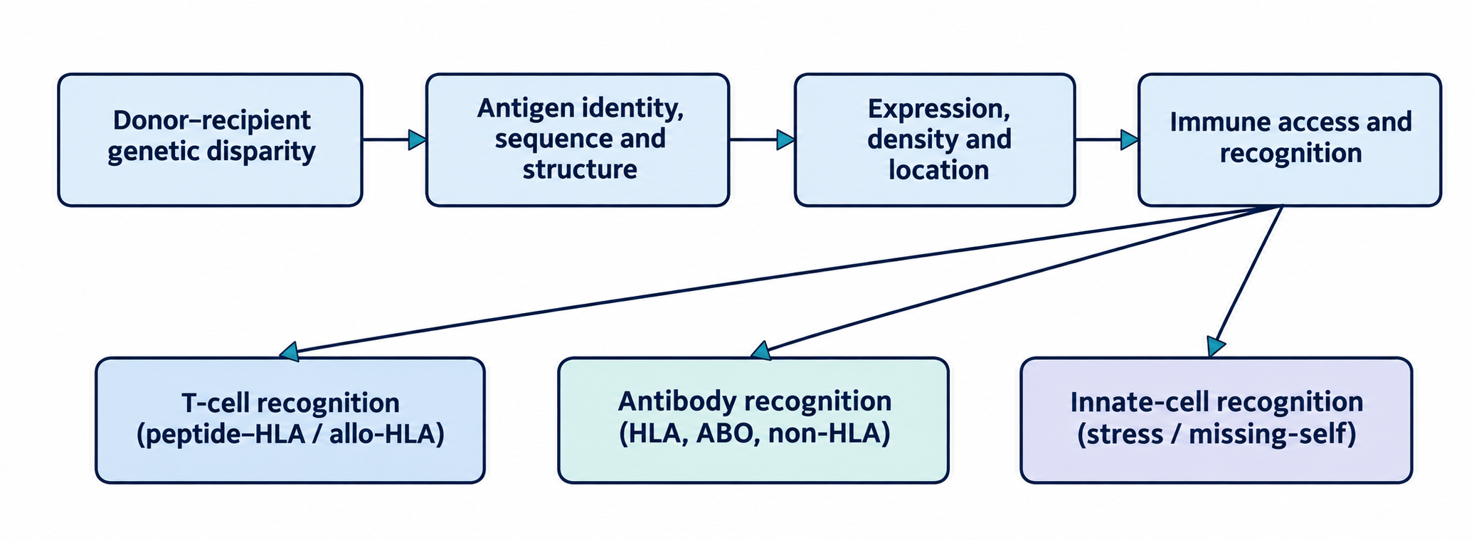
Graft compatibility is not a single molecular property. It is the outcome of an interaction between donor–recipient genetic disparity, the antigenic molecules or peptides generated from that disparity, the distribution and abundance of those targets, and the immune repertoire and physiological state of the recipient or donor-derived immune system. Classical HLA molecules are the dominant transplantation antigens because they are highly polymorphic, widely expressed, and central to peptide presentation and T-cell recognition. However, the antigenic landscape extends beyond HLA to include minor histocompatibility antigens, ABO carbohydrates, non-classical MHC molecules, MICA/MICB, polymorphic endothelial or epithelial proteins, and tissue-associated structures exposed or modified during injury. The same genetic difference can have distinct consequences depending on the transplant setting. In solid-organ transplantation, recipient immunity encounters donor antigens on graft endothelium, parenchymal cells and passenger leukocytes. In allogeneic HCT, donor-derived immune cells develop or mature in the recipient and can recognise recipient polymorphic peptides and other antigens. Thus, donor–recipient disparity can contribute to graft-versus-host disease (GVHD), graft-versus-leukaemia (GVL) activity, graft rejection or graft failure. HLA matching is therefore a risk-reduction strategy, not a guarantee of complete immunological identity.
2.2.1 Self, Non-Self and Altered-Self Antigen Recognition in Transplantation
Self and non-self are foundational immunological concepts, but transplantation exposes the limitations of a simple binary model. A recipient can recognise intact donor HLA as allogeneic, recognise donor-derived peptides presented by recipient HLA, or recognise polymorphic peptides from donor or recipient proteins. Antibodies may bind conformational epitopes on intact donor molecules, while innate lymphocytes may respond to altered expression of self-MHC or stress-induced ligands. The immune system therefore detects molecular patterns and context rather than applying a single universal self/non-self rule.
Central tolerance removes or edits many developing lymphocytes with strong reactivity to self-antigens, while peripheral tolerance mechanisms restrain potentially autoreactive or cross-reactive cells. These mechanisms do not eliminate all T-cell reactivity to allogeneic MHC. The alloreactive repertoire is unusually broad because T-cell receptors selected to recognise self-peptide–self-MHC can cross-react with structurally different allogeneic peptide–MHC complexes. The high frequency of alloreactive T cells is one reason that HLA disparity can provoke strong responses. Detailed structural and cellular pathways are addressed in Module 3.
Non-self recognition is not restricted to intact foreign proteins. In indirect recognition, recipient antigen-presenting cells process donor proteins and display donor-derived peptides on recipient HLA. Minor histocompatibility antigens provide another example: the HLA molecule may be shared, but the peptide sequence differs. The immune system recognises the altered peptide–HLA surface as a distinct antigenic structure. Conversely, a polymorphism that does not alter an expressed protein, peptide processing, binding or immune accessibility may have little or no immunological consequence.
Altered-self recognition is particularly relevant to innate immunity and tissue injury. Stress can change the expression of ligands for activating or inhibitory receptors, modify HLA expression, expose intracellular molecules, or generate neoepitopes through oxidation, cleavage, post-translational modification or cell death. MICA and MICB are stress-inducible MHC class I-related molecules that can engage NKG2D on NK cells and subsets of T cells. Their biology illustrates how tissue stress and antigen expression can alter immune recognition without a conventional donor-specific HLA mismatch.
Transplantation also creates an unusual relationship between tolerance and immunity. The recipient is expected to tolerate donor-derived tissue while retaining protective immunity against pathogens and malignancy. In HCT, donor-derived immune cells must establish tolerance to recipient tissues while preserving GVL activity. The immunological goal is therefore not simply to suppress recognition of all foreign structures; it is to shape the specificity, magnitude, location and effector function of the response.
2.2.2 Definition and Classification of Transplant Antigens
Transplantation antigens are molecular structures that can be recognised by the immune system of a genetically distinct donor or recipient and thereby influence graft acceptance, injury or rejection. In the strict immunogenetic sense, a transplantation antigen is not simply any molecule present in a graft. It is a structure whose inherited or acquired variation, expression or presentation creates a target for an immune response in the transplant setting. The term includes classical histocompatibility antigens, but the functional antigenic landscape is broader and includes carbohydrate antigens, polymorphic peptides, non-HLA proteins and stress-associated ligands.
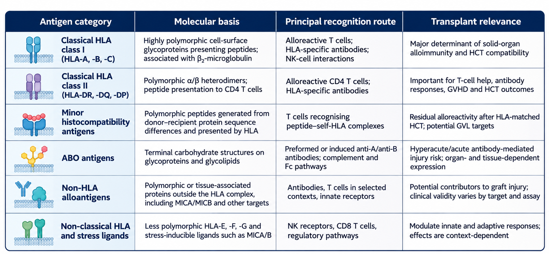
Major histocompatibility antigens are encoded within the MHC. In humans, the MHC is the HLA complex on chromosome 6p21.3. Classical HLA class I and class II molecules are highly polymorphic and are central to antigen presentation. Their importance reflects both their direct recognition by alloreactive T cells and their capacity to present peptides derived from donor or recipient proteins. HLA class I molecules are expressed on most nucleated cells, whereas class II expression is concentrated on professional antigen-presenting cells but can be induced in other cell types, including endothelial and epithelial compartments under inflammatory conditions.
Minor histocompatibility antigens (MiHAs) are polymorphic peptides derived from cellular proteins. They are generated when donor and recipient differ at a coding polymorphism, such as a single-nucleotide variant, insertion/deletion or other sequence variation, and the resulting peptide is presented by an HLA molecule shared between donor and recipient. Thus, MiHA disparity can remain even when donor and recipient are HLA matched. Their biological effects depend strongly on the tissue distribution of the source protein and the HLA restriction of the responding T cell.
ABO antigens are carbohydrate structures rather than HLA-like peptide-presenting molecules. A and B determinants are expressed on erythrocytes and, depending on tissue and glycan structure, on vascular endothelium and other cells. Naturally occurring anti-A and anti-B antibodies can bind incompatible graft structures and activate complement or Fc-mediated effector pathways. The distribution and density of ABO structures are not identical across tissues, which helps explain why ABO incompatibility has different implications across organ types and transplant protocols.
Non-HLA alloantigens include polymorphic proteins outside the MHC, tissue-associated antigens and molecules that become immunologically accessible after injury. Examples discussed in transplantation include MICA/MICB, angiotensin II type 1 receptor (AT1R), endothelial-associated targets, vimentin, collagen and other structural or intracellular proteins. Evidence differs substantially across targets: biological plausibility, antibody detection and association with injury do not automatically establish a validated diagnostic biomarker or a universal clinical indication for testing.
Other polymorphic cell-surface systems include human neutrophil antigens (HNA) and human platelet antigens (HPA). HNAs are carried by neutrophil-associated glycoproteins (including FcγRIIIb/CD16b, CD177, CD11/18-associated structures and other neutrophil proteins), whereas HPAs are polymorphic determinants on platelet glycoproteins such as GPIIb/IIIa, GPIb/IX/V, GPIa/IIa and CD109. These systems are important in transfusion medicine and alloimmune cytopenias, including transfusion-related acute lung injury, neonatal alloimmune neutropenia, fetal/neonatal alloimmune thrombocytopenia and platelet transfusion refractoriness. Some HPA-bearing glycoproteins are also expressed on endothelial or other cell types, but HNA/HPA incompatibility is not generally treated as a primary HLA-equivalent determinant of routine solid-organ compatibility.
Classification is therefore best performed along several axes: genetic location (HLA versus non-HLA), molecular form (protein, peptide, carbohydrate or lipid-associated structure), recognition route (T-cell, antibody or innate receptor), tissue distribution (haematopoietic-restricted, endothelial, epithelial or ubiquitous), and transplant context (solid organ versus HCT). These dimensions are more informative than treating all transplantation antigens as a single category. Importantly, not every clinically discussed non-HLA target is necessarily a donor-polymorphic alloantigen. Some antibodies described in transplantation are directed against self-proteins or injury-associated structures. Such reactivity may arise through autoimmunity, altered antigen availability, inflammation or exposure of previously sequestered structures; the mechanism is not automatically an alloimmune response to a donor-specific sequence variant. These targets should therefore be described as non-HLA antibody targets or injury-associated autoantigens unless donor–recipient polymorphism and allo-specificity have been demonstrated. This distinction matters when interpreting assay positivity and causal claims.
2.2.3 Major Histocompatibility Antigens (HLA) and Their Role in Graft Compatibility
The MHC is a gene-dense, highly polymorphic region of the genome. In humans, the HLA complex occupies the short arm of chromosome 6 and contains classical class I and class II loci, non-classical HLA genes and numerous immune-related genes. The classical class I loci HLA-A, HLA-B and HLA-C encode heavy chains that associate with β2-microglobulin. The classical class II molecules HLA-DR, HLA-DQ and HLA-DP are heterodimers composed of polymorphic α and β chains. Their peptide-binding domains contain much of the polymorphic variation that shapes peptide repertoire and T-cell recognition.
HLA class I molecules generally present peptides derived from intracellular proteins to CD8-positive T cells. The heavy chain contains three extracellular domains (α1, α2, α3), a transmembrane region and a cytoplasmic tail. The heavy chain associates non-covalently with β2-microglobulin which stabilises the complex on the cell surface. Peptides are commonly generated by proteasomal degradation, transported into the endoplasmic reticulum by TAP proteins and loaded onto nascent HLA class I molecules. The detailed mechanism of antigen presentation is reserved for Module 3.
A related but conceptually distinct layer is the interaction between HLA class I ligands and killer-cell immunoglobulin-like receptors (KIR). Polymorphic HLA class I molecules provide ligands for inhibitory or activating KIR; inhibitory receptor engagement restrains NK-cell activity, whereas inadequate inhibitory input can contribute to 'missing-self' recognition. Its relevance is particularly discussed in HCT and GVL biology, but clinical donor-selection effects are context-dependent and not uniformly established.
HLA class II molecules present peptides, often derived from extracellular or endosomal proteins to CD4-positive T cells. They are expressed constitutively by professional antigen-presenting cells, including dendritic cells, B cells and macrophages. Inflammatory cytokines, particularly interferon-γ, can induce class II expression in other cell types like epithelial cells through transcriptional control involving CIITA. Class II disparity can therefore influence both direct T-cell recognition and the recipient's capacity to generate helper responses that support alloantibody formation. The detailed mechanism of antigen presentation is reserved for Module 3.
HLA molecules are codominantly expressed: alleles inherited from both parents can be expressed. This means that an individual generally expresses multiple HLA-A, -B and -C class I allotypes and multiple class II heterodimers, subject to locus-specific gene structure and expression. Classical HLA polymorphism has two major consequences. First, amino-acid differences can alter the shape and chemistry of the peptide-binding groove, affecting which peptides can be presented. Second, polymorphic residues can form epitopes recognised by antibodies or contribute to structural surfaces recognised by T cells. An HLA mismatch is therefore not merely a different allele label: it can represent a difference in peptide repertoire, surface conformation and immune recognition potential. However, not all mismatches have equivalent biological effects, and the clinical importance depends on locus, allele, expression, antibody status, transplant type and prophylaxis.
Non-classical HLA molecules provide additional layers of regulation. HLA-E presents a restricted peptide repertoire and interacts with CD94/NKG2 receptors on NK cells and subsets of T cells. HLA-G has immunoregulatory functions and restricted expression patterns, particularly at maternal–fetal interfaces, although its roles in transplantation remain context-dependent. HLA-F has distinct expression and receptor interactions. MICA and MICB are MHC class I-related molecules but are not classical peptide-presenting HLA molecules; they function as stress ligands and are polymorphic.
The MHC also contains a class III region, but the term 'class III' does not refer to a third group of classical peptide-presenting HLA molecules. It includes genes encoding immune and inflammatory proteins, among them complement components (C2, C4 and factor B), tumour-necrosis-factor-family cytokines and other immune-related products. These genes can influence inflammatory and complement biology and may be inherited in linkage disequilibrium with HLA alleles, but they should not be conflated with HLA-A/B/C or HLA-DR/DQ/DP antigens.
2.2.4 Minor Histocompatibility Antigens (miHA) and Non-HLA Alloantigens
MiHAs are peptide antigens derived from polymorphic proteins encoded outside the HLA loci. A MiHA becomes relevant when a donor and recipient differ at a polymorphic site, the recipient or donor expresses the relevant source protein, the variant peptide can be processed and presented by an HLA molecule, and a T-cell repertoire capable of recognising that peptide–HLA complex is present. The term 'minor' refers historically to their generally weaker or more restricted effects compared with major MHC disparities; but it does not mean that their clinical consequences are trivial. In HLA-matched HCT, donor T cells can recognise recipient MiHAs presented by shared HLA molecules. If the source protein is broadly expressed, the target can be present in both haematopoietic and non-haematopoietic tissues, potentially contributing to GVHD as well as GVL. If expression is restricted predominantly to haematopoietic cells, the response may preferentially target residual recipient haematopoiesis and malignant cells while sparing many non-haematopoietic tissues. This tissue-restricted biology is a central rationale for MiHA-directed immunotherapy.
Recent systematic work has expanded the catalogue and analytical identification of MiHAs. HA-1, HA-2, HA-3, HA-4, HA-8 and H-Y are particularly well-characterised examples. HA-1 is a well-studied example of a haematopoietic-restricted MiHA presented by HLA-A*02:01. Its immunogenic and non-immunogenic forms arise from a polymorphism in HMHA1/ARHGAP45. HA-2, encoded by MYO1G and also presented by HLA-A*02:01, is another example. These targets illustrate that antigen-specific donor T-cell responses can potentially be directed toward malignant recipient haematopoietic cells after HCT. Translation requires careful genotyping of both donor and recipient, confirmation of HLA restriction, expression in target cells and avoidance of off-target recognition.
Unlike MiHAs which are presented by HLA molecules to T cells, non-HLA alloantigens are a broader set of polymorphic or tissue-associated targets. MICA and MICB are encoded within the MHC region but are not classical HLA class I molecules. They are stress-inducible and can be expressed independently on endothelial and epithelial cells. Antibodies against MICA have been associated with graft injury in some settings, but the interpretation of non-HLA antibody testing is limited by assay standardisation, antigen presentation, specificity assignment and variable evidence for clinical utility. Other reported targets include AT1R, endothelin receptor type A, vimentin, collagen and perlecan-associated structures. Their relevance should be considered individually rather than treated as one validated antibody category. Mechanistically, MICA and MICB should not be described as conventional HLA class I antigen-presenting molecules. They do not associate with β2-microglobulin and do not form a peptide-binding groove. Instead, they act primarily as stress-inducible ligands for NKG2D, expressed by NK cells and subsets of T cells. Their expression can rise on endothelial and epithelial cells in response to cellular stress, infection, tissue damage or malignant transformation; soluble shedding or extracellular-vesicle release can also alter NKG2D signalling. Anti-MICA antibodies have been associated with graft injury in selected settings, but their interpretation requires consideration of target expression, assay design, antibody specificity and clinicopathological evidence.
HNA and HPA systems belong in the broader inventory of non-HLA polymorphic antigen systems, but their clinical setting should be stated precisely. HNA alloantibodies can cause neutropenia or transfusion-related lung injury, while HPA alloantibodies are important in fetal/neonatal alloimmune thrombocytopenia and platelet refractoriness. Platelets also carry HLA class I and ABH determinants. These antigens can become relevant to transfusion support around transplantation and, in selected circumstances, to immune complications, but they are distinct from the principal HLA and MiHA pathways driving conventional graft histocompatibility. The importance of these non-HLA alloantigens is that pre-existing alloantibodies can target them and result in clearance of cells bearing them on their surface.
Non-HLA alloantigens can also include polymorphic proteins that are not routinely measured in standard HLA laboratories. Their potential immunogenicity depends on whether they are accessible to antibodies, processed and presented to T cells, or recognised by innate receptors. A target located intracellularly may become accessible following cell injury or may generate peptides for HLA presentation without being directly accessible to circulating antibody. Distinguishing these routes is essential when evaluating proposed mechanisms of graft injury.
2.2.5 Antigen Expression and Availability
Antigen expression determines whether a genetically encoded target is available for immune recognition. Classical HLA class I molecules are expressed on nearly all nucleated cells, although abundance varies among loci, alleles, cell types and activation states. HLA class II molecules are constitutively expressed on professional antigen-presenting cells and can be induced on endothelial, epithelial and other cells during inflammation. These patterns mean that the same HLA disparity may be encountered across multiple graft compartments and can become more visible during inflammatory injury.
ABO antigen expression is tissue-dependent. ABO carbohydrate structures are found on erythrocytes and on endothelial or epithelial surfaces in a tissue. The precise glycan structures and density can differ between cell types, so RBC phenotype alone does not fully describe tissue expression. This distinction is relevant when considering ABO-incompatible transplantation and the biological basis of accommodation or antibody-mediated injury. The molecular basis of this tissue dependence is the glycan backbone on which the terminal A, B or H determinants are displayed. ABH structures occur on different precursor chains (commonly designated type I–IV), and the relative abundance of these structures differs between erythrocytes, vascular endothelium and epithelial tissues. In a study of heart and spleen tissue, endothelial ABH expression was predominantly type-II based, whereas erythrocytes displayed a broader and quantitatively different pattern. Thus, red-cell agglutination titres provide an operational measure of circulating anti-ABO reactivity but do not perfectly quantify antigen abundance or glycan context on every graft tissue.
Endothelial cells are particularly important in vascularised solid-organ transplantation because they form the interface between recipient blood and donor tissue. They express HLA class I and can express HLA class II under inflammatory conditions. They are directly exposed to circulating antibodies, complement and leukocytes. Consequently, endothelial antigen expression is central to antibody-mediated injury and to the interpretation of non-HLA endothelial targets.
Parenchymal and epithelial compartments differ by organ. Renal tubular epithelial cells, hepatocytes, biliary epithelium, airway epithelium, cardiomyocytes and intestinal epithelial cells have distinct basal and inducible antigen-expression patterns. Local cytokines, infection, ischaemia-reperfusion injury and tissue remodelling can alter HLA expression and antigen processing. An antigen that is weakly expressed in resting tissue may become more abundant after inflammatory stimulation.
Haematopoietic cells express HLA and can serve as targets of donor- or recipient-derived immune responses. In HCT, the cellular composition of the graft and the recipient's haematopoietic and non-haematopoietic compartments change over time as donor engraftment proceeds. Recipient antigen-presenting cells may persist during early phases, while donor-derived antigen-presenting cells progressively contribute to immune reconstitution. This dynamic cellular origin affects which antigens are presented and which tissues are vulnerable to alloreactivity.
Non-classical HLA molecules and stress ligands have more restricted or inducible distributions. MICA/MICB can be upregulated by cellular stress and are expressed in selected epithelial and endothelial settings. HLA-G has restricted expression and immunoregulatory properties. HLA-E expression is linked to the availability of leader-sequence peptides from other HLA class I molecules. These molecules influence NK-cell and other immune-cell responses and should be considered as regulators of the antigenic environment, not simply as additional classical HLA loci.
2.2.6 Antigen Density, Tissue Distribution, and Immunological Accessibility
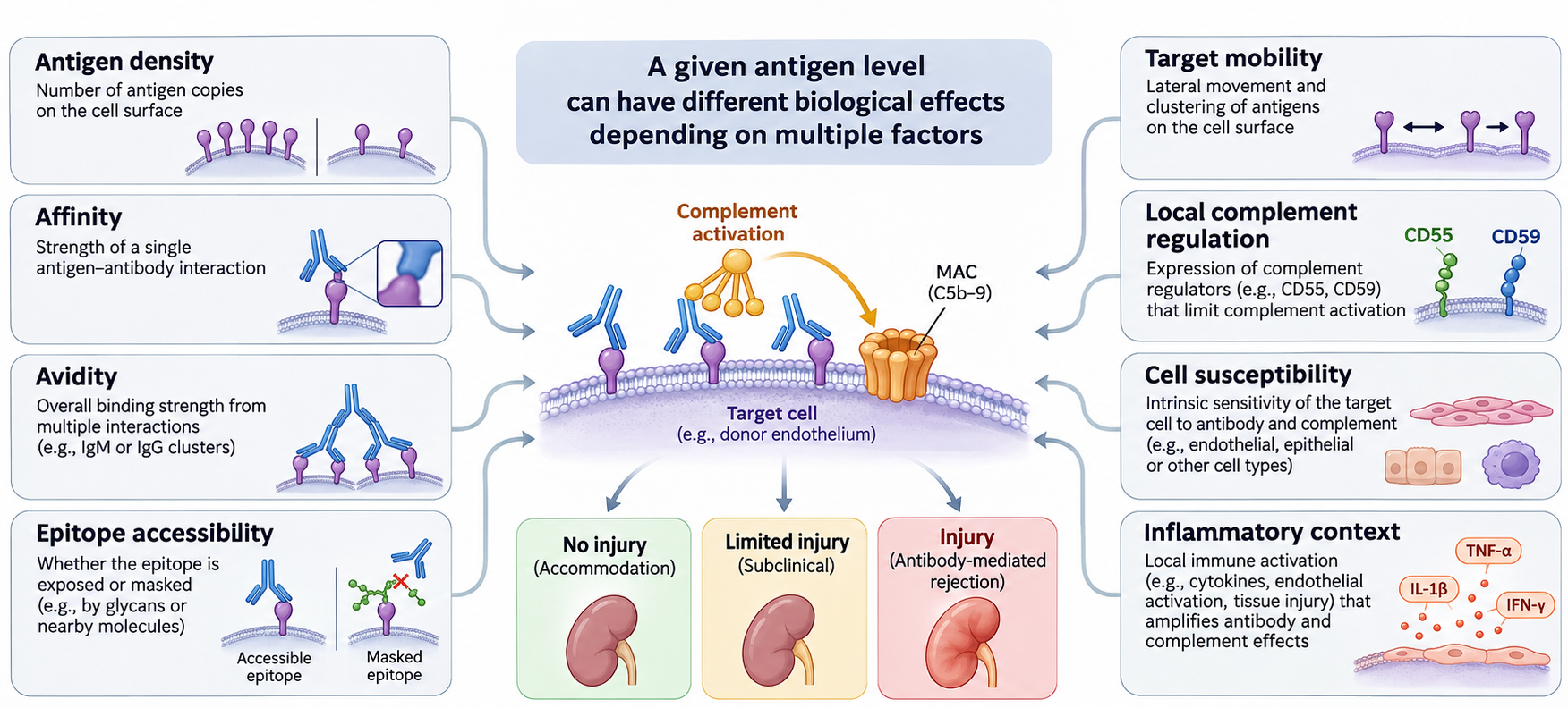
Affinity, avidity, epitope accessibility, target mobility, local complement regulation, cell susceptibility and inflammatory context all modify the biological effect of a given antigen level.
Antigen density refers to the abundance of a target antigen at the cell surface or within a relevant tissue compartment. It can influence the probability of antibody binding, receptor engagement, immune synapse formation and effector activation. HLA expression is not uniform. Expression varies between HLA loci, alleles, tissues and activation states. HLA-A and HLA-B generally show higher surface expression than HLA-C, while HLA-DQ and HLA-DP expression can differ substantially by cell type and regulatory context. Allele-specific promoter variants, transcriptional regulators, alternative splicing, epigenetic modifications and microRNA-mediated effects can alter expression. These differences can affect the amount of target available to antibodies or T cells, but should not be converted into a fixed clinical hierarchy without specifying the tissue, assay and transplant context.
Inflammatory cytokines can change antigen abundance. Interferon-γ can increase class I and class II expression, while type I interferons and other inflammatory signals can also modulate class I pathways. This means that an antigen's expression at the time of pretransplant typing may not represent its abundance after transplantation, particularly in the presence of ischaemia-reperfusion injury, infection or rejection. The graft's antigenic phenotype is dynamic rather than fixed.
Accessibility describes whether an immune effector can physically encounter the target. Surface-expressed HLA and ABO structures are accessible to circulating antibodies. Intracellular proteins are not directly accessible to antibody in intact cells, but their peptides can be presented by HLA molecules to T cells (miHA). Tissue architecture, vascular perfusion, extracellular matrix, cell polarity and local inflammation influence access. An antigen may be present but immunologically sequestered until tissue injury exposes it.
Tissue and cell distribution determines the consequences of recognition. A broadly expressed MiHA can be present on both haematopoietic and non-haematopoietic tissues, increasing the potential for GVHD. A haematopoietic-restricted MiHA may preferentially direct donor T cells toward residual recipient blood cells or malignant cells, offering a route to GVL with reduced non-haematopoietic tissue injury. The distinction is not absolute: expression can be heterogeneous, and inflammatory states may alter tissue expression.
Antigen density and tissue distribution also interact with antibody properties. A strong antibody signal against a low-density antigen may not have the same biological meaning as a similar signal against a highly expressed target. Conversely, a low-abundance antigen can be important if the antibody has high affinity, binds an accessible functional epitope or activates potent effector pathways. This is one reason why antibody assay intensity cannot be interpreted as a direct quantitative measure of graft injury risk without considering antigen expression and assay characteristics.
2.2.7 Determinants of Graft Compatibility
Overall, donor-recipient genetic compatibility as well as immune repertoire are major determinants of graft combability.
Donor–recipient genetic disparity can occur at multiple levels: whole haplotypes (e.g. differences in inherited HLA-A, -B, -C, -DRB1 and -DQB1 haplotypes), HLA alleles (e.g. HLA-A*02:01 versus HLA-A*02:05), individual amino-acid residues (e.g. a mismatch at position 116 of an HLA-B molecule), non-HLA coding variants (e.g. HMHA1 polymorphisms encoding the HA-1 minor histocompatibility antigen), regulatory variants (e.g. polymorphisms in the HLA-C promoter affecting gene expression), structural variants (e.g. deletion of an HLA gene) and copy-number differences (e.g. variation in C4A or C4B gene copy number within the MHC region). Each level captures a different aspect of biological similarity.
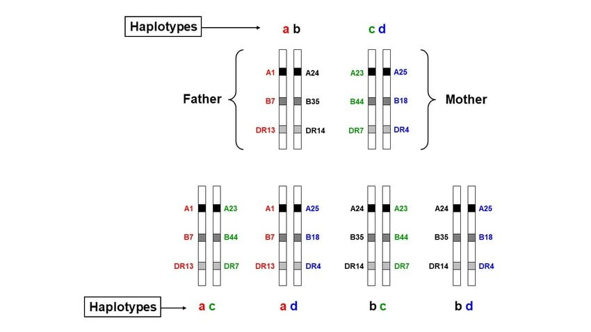
A haplotype is a group of alleles at different genetic loci that are located physically on the same chromosome and are inherited together from one parent. HLA genes are inherited as haplotypes. Because neighboring HLA genes are physically close to one another, recombination between them occurs relatively infrequently. As a result, certain combinations of HLA alleles at neighbouring loci are transmitted together across generations more frequently than would be expected by chance. This phenomenon is called linkage disequilibrium (LD). Indeed, the relationship is that haplotypes are the physical combinations of alleles on individual chromosomes, whereas linkage disequilibrium describes how frequently particular allele combinations occur together across a population. Accordingly, a related donor may share one or both parental haplotypes with the recipient. Unrelated individuals however can share common alleles but differ across other loci or haplotypes. For a simple family in which both parents carry two distinguishable HLA haplotypes (let’s ignore recombination; F1 F2 and M1 M2), each child potentially inherit four different combinations (F1M1, F1M2, F2M1, F2M2). Same rule applies to every child. If we compare siblings, there are 25%, 50% and 25% chance that two siblings sharing zero, one, or two HLA haplotypes. These Mendelian probabilities are useful for understanding related-donor searches, but they are not a substitute for actual donor and recipient typing.
An HLA mismatch can be described at antigen level, allele level, amino-acid level or structural epitope level. Antigen-level comparisons reflect broad serologically defined specificities (antibody-based testing). Allele-level typing distinguishes sequence-defined alleles (NGS methods). Amino-acid mismatch analyses count or characterise residue differences, often within antigen-recognition domains. Eplet or epitope approaches attempt to describe antibody-accessible structural configurations. These approaches are related but not interchangeable, and their predictive performance differs across clinical settings.
As discussed, donor–recipient differences outside HLA can cause genetic disparity. Polymorphic miHA peptides, ABO carbohydrate antigen and non-HLA polymorphisms contribute to genetic differences.
Antigen expression, tissue distribution, antigen density, and immunological accessibility across donor organs and tissues represent additional dimensions of graft compatibility. While we have greater capacity to investigate and manipulate some of these variables, differences peptide-binding groove, peptide pools, gene expression, promoter polymorphisms, transcription-factor binding, methylation, alternative splicing, microRNA regulation and protein turnover, peptide repertoires and immune receptor landscapes represent dimensions of alloreactivity that remain less understood and more difficult to control. Such functional differences are important because even when the HLA molecules themselves are shared between donor and recipient, donor–recipient polymorphisms can create different peptides pools that consequently could result in allo-immunity. Histocompatibility is therefore a composite property rather than a single genotype score.
2.2.8 Histocompatibility in Solid-Organ Transplantation
In solid-organ transplantation, the graft retains donor-derived endothelial, epithelial, parenchymal and resident immune cells. The recipient's immune system encounters these donor structures after reperfusion and can mount cellular and humoral responses. HLA disparity is a major determinant of alloimmune risk, but the effect of mismatch varies by organ, locus, sensitisation status, immunosuppressive regimen and the clinical endpoint being considered. Kidney transplantation has a particularly developed evidence base for HLA matching and antibody assessment; other organs differ in the relative contribution of HLA, ABO, non-HLA antibodies and organ-specific immunobiology.
Preformed recipient HLA antibodies can bind donor HLA on graft endothelium and contribute to early antibody-mediated injury. De novo alloantibodies can develop after transplantation through T-cell-dependent and other B-cell pathways. Donor-specific antibody is defined by its specificity for HLA expressed by the donor, but the presence of DSA does not by itself establish tissue injury. Antibody strength, persistence, complement-binding properties, target expression, endothelial susceptibility, histology and graft function all influence interpretation.
ABO compatibility is an additional major consideration for many vascularised organs because anti-A or anti-B antibodies can bind donor vascular structures. ABO-incompatible transplantation is possible in selected settings with specialised protocols, but the biological risk and clinical approach differ by organ, recipient age, antibody titre, graft characteristics and centre experience. ABO compatibility should not be confused with HLA compatibility: they involve different antigen chemistries and different immune recognition pathways.
Non-HLA targets may contribute to graft injury, particularly when endothelial or epithelial cells are stressed. MICA/MICB antibodies and antibodies to AT1R or other tissue-associated targets have been associated with selected clinical phenotypes. However, non-HLA antibody assays vary in antigen source, conformation, threshold and validation. Association with adverse outcome is not sufficient to justify treating every detected antibody as pathogenic or to apply a uniform testing algorithm.
Organ-specific expression patterns influence histocompatibility. The kidney has a large endothelial and tubular epithelial surface; the lung contains extensive airway and alveolar epithelial compartments; the heart has vascular endothelium and cardiomyocytes; the liver has specialised sinusoidal endothelium, hepatocytes and biliary epithelium; and the intestine contains a dense immune and epithelial interface. These differences shape antigen accessibility, local immune activation and tissue injury patterns.
Histocompatibility in solid-organ transplantation is therefore a risk continuum. A donor–recipient pair may be HLA matched but ABO incompatible, HLA mismatched but without detectable donor-specific antibody, or genetically similar yet exposed to non-HLA targets or tissue injury. The clinically relevant question is not simply whether a mismatch exists, but whether the mismatch can be recognised by the recipient's immune system and whether that recognition is likely to damage the graft under the planned clinical conditions.

The clinical weight assigned to HLA matching is not identical across organs. The biology of the graft, the urgency of allocation, the ability to perform prospective testing, the prevalence and consequences of preformed donor-specific antibody, and organ-specific tolerance or resistance to antibody injury all influence how histocompatibility data are used. The following comparison is intentionally qualitative; allocation policies and crossmatch requirements vary by jurisdiction and transplant programme.
| Organ / graft | Compatibility considerations | Interpretive caution |
|---|---|---|
| Kidney | The most established clinical evidence base for HLA matching; HLA mismatch burden, preformed/de novo DSA and crossmatch status can influence rejection and long-term graft outcomes. ABO compatibility is also important, although selected ABO-incompatible programmes are established. | The relative priority of HLA matching depends on living/deceased donor context, allocation system, recipient sensitisation and centre policy. |
| Heart | Routine allocation is not generally based on direct HLA matching in many systems; antibody screening and crossmatch are particularly important for sensitised candidates. DSA and antibody-mediated injury remain clinically important. | Absence of routine HLA matching in allocation does not mean HLA antibodies are biologically irrelevant. |
| Lung | Allocation usually places greater weight on urgency and physiological matching than on direct HLA matching; pretransplant antibodies, unacceptable antigens and crossmatch strategy are central in sensitised candidates. | The lung is highly immunologically exposed; DSA and antibody-mediated injury can be consequential despite the absence of routine allele matching. |
| Liver | HLA matching is not routinely used as a principal allocation criterion. Liver grafts show distinctive immunoregulatory and antibody-handling properties, but ABO compatibility and selected DSA-related risks remain relevant. | Do not describe the liver as immunologically privileged or immune to antibody injury; tolerance is relative and phenotype-dependent. |
| Pancreas / islet | HLA and DSA are relevant to alloimmune risk, and ABO compatibility and recipient sensitisation are considered; clinical emphasis varies by whole-organ versus islet procedure. | Evidence and allocation practices differ from kidney transplantation; avoid importing kidney matching rules without qualification. |
| Intestine / multivisceral and VCA | Graft immune-cell content, epithelial/skin exposure and tissue-specific antigen expression create distinctive alloimmune risks; HLA, ABO and antibody assessments are interpreted alongside graft-specific factors. | These grafts have distinct immunobiology; their compatibility cannot be reduced to a single HLA mismatch count. |
2.2.9 Histocompatibility in Allogeneic Haematopoietic Cell Transplantation
Allogeneic HCT differs fundamentally from solid-organ transplantation because the graft contains haematopoietic stem and progenitor cells together with variable numbers of mature immune cells. After conditioning, donor-derived haematopoiesis becomes established and the donor immune system can recognise recipient tissues. Histocompatibility therefore involves bidirectional immune risk: recipient immunity can reject donor cells, while donor immunity can attack recipient tissues or malignant cells.
HLA matching remains a major determinant of HCT outcome, but its definition must state the loci and typing resolution. The 2026 EBMT Practice Harmonisation and Guidelines Committee recommends that patients and potential donors have, at minimum, high-resolution typing at HLA-A, -B, -C, -DRB1, -DQB1 and -DPB1. Where feasible, the preferred approach is ultra-high-resolution typing for these six loci, with additional high-resolution typing of HLA-DQA1, -DPA1 and -DRB3/4/5 (11 loci in total), particularly when donor-specific antibody assessment may be relevant. EBMT commonly uses a 10/10 definition based on HLA-A, -B, -C, -DRB1 and -DQB1; CIBMTR’s commonly used 8/8 definition excludes DQB1. The numerical label must therefore be accompanied by the loci included.
This expanded typing approach helps identify allele-level differences, null or differentially expressed alleles, and recombination-related complications that may be missed by narrower panels. For a matched sibling, family-based haplotype assignment can help confirm identity by descent and detect uncommon recombination. A second, non-haematopoietic sample may be needed when blood-derived typing could be affected by malignancy-associated loss of heterozygosity, allele dropout or prior allogeneic transplantation.
Donor-specific anti-HLA antibody testing should be performed before selecting a mismatched donor, because recipient antibodies directed against donor HLA can impair engraftment. The relative importance of mismatch models also depends on the planned GvHD prophylaxis platform. Much of the evidence supporting permissive mismatch models derives from calcineurin-inhibitor-based, non-PTCy settings; the evidence base under post-transplant cyclophosphamide (PTCy) is less mature. Donor age, time-to-transplant, disease urgency, graft source, DSA and prophylaxis should therefore be considered together rather than applying a rigid universal donor hierarchy.
Mismatch directionality matters. In the graft-versus-host direction, donor T cells can recognise recipient antigens not present in the donor. In the host-versus-graft direction, recipient immune cells can recognise donor antigens and contribute to rejection or graft failure. A bidirectional mismatch can create both risks. Haploidentical transplantation is characterised by one shared and one unshared HLA haplotype; the mismatch burden and direction should be interpreted in relation to the conditioning and GVHD prophylaxis platform.
MiHAs remain relevant in HLA-matched HCT. Donor T cells can recognise recipient polymorphic peptides presented by shared HLA. Broadly expressed MiHAs can contribute to GVHD and GVL, whereas haematopoietic-restricted MiHAs may support more selective GVL. H-Y antigens can become relevant in sex-mismatched transplantation. The source tissue and cellular origin of the antigen are therefore critical when considering the balance between antitumour activity and GVHD.
Modern GVHD prophylaxis has changed the interpretation of HLA mismatch. Post-transplant cyclophosphamide and other contemporary strategies have enabled successful transplantation across mismatched donor sources, expanding access for patients without a fully matched donor. This does not mean that HLA disparity is biologically irrelevant; rather, the clinical impact of a mismatch is modified by prophylaxis, donor age, donor-specific antibodies, timing, graft source and disease context. Recent EBMT recommendations emphasise that donor selection should be integrated and contextual rather than based on a single rigid hierarchy.
Donor-specific HLA antibodies are especially important in mismatched HCT because recipient antibodies directed against donor HLA can impair engraftment. Antibody testing should therefore be considered when selecting mismatched donors, with the target specificity interpreted against the actual donor HLA genotype. This is a distinct risk from donor T-cell alloreactivity against recipient tissues. Both directions must be evaluated.
Donor selection also considers non-HLA factors, including donor age, availability, urgency, graft source, CMV status in selected contexts, ABO compatibility and other donor characteristics. These variables do not replace HLA assessment, but contemporary recommendations recognise that the optimal donor is a composite decision. In particular, a timely alternative donor transplant may be preferable to a prolonged search for a theoretically ideal match when disease urgency is high.
KIR–HLA interactions add an additional layer to HCT immunobiology. Donor NK cells express inhibitory and activating KIR receptors that interact with recipient HLA class I ligands; the resulting balance can alter NK-cell education, inhibition and potential GVL activity. The ligand–receptor framework is biologically established, but the clinical benefit of selecting donors by individual KIR/HLA combinations has been inconsistent across studies. A large 2024 analysis did not find consistent outcome associations for individual donor-KIR/recipient-HLA interactions in HLA-matched unrelated-donor HCT for AML, underscoring the need to avoid treating KIR genotype as a validated stand-alone donor-ranking rule.
2.2.10 Biological Limits of HLA Matching and the Concept of Residual Alloreactivity
HLA matching substantially reduces major histocompatibility disparity, but it does not create complete donor–recipient immunological identity. First, conventional matching examines selected HLA loci and defined sequence resolution; it does not establish identity across every MHC gene, regulatory region or non-HLA locus. Second, HLA-matched individuals can differ at polymorphic proteins that generate MiHAs. Third, ABO, MICA/MICB and other non-HLA systems can remain disparate. Fourth, allele-specific expression and tissue-specific antigen abundance can differ despite a nominal match. Therefore, residual alloreactivity can arise.
Residual alloreactivity arises through several routes. Donor or recipient T cells may recognise MiHA peptides presented by shared HLA. Recipient antibodies may target donor HLA or non-HLA antigens. NK cells may respond to differences in inhibitory ligand availability, activating ligands or stress-associated expression. Inflammatory injury can increase HLA expression and expose otherwise inaccessible targets. Microbiota, infection, tissue damage and cytokine environments can modify the threshold for immune activation.
Even HLA-identical sibling transplantation does not eliminate GVHD. Minor antigen disparities, sex-linked antigens, age-related or acquired cellular changes, and tissue-specific inflammatory states can sustain donor T-cell responses. Conversely, HLA-mismatched transplantation can be successful under appropriate clinical conditions, especially when donor selection, antibody assessment, conditioning and GVHD prophylaxis are optimised. The relationship between matching and outcome is therefore probabilistic, not deterministic.
Residual alloreactivity exposes the limitations of purely genotype-based prediction. A mismatch may not be immunogenic if the antigen is not expressed, not processed, not presented by the relevant HLA molecule or not recognised by the available immune repertoire. Conversely, a small number of highly consequential differences may dominate risk if they are expressed at high density in accessible tissue and recognised by pre-existing antibodies or high-frequency T-cell clones.
Precision compatibility assessment therefore requires integration of multiple data layers: high-resolution HLA genotype; donor-specific antibody profile; ABO status; relevant non-HLA targets where validated; expression and tissue distribution; transplant type and directionality; donor and recipient clinical variables; and the planned immunosuppressive or GVHD prophylaxis strategy. Emerging genomic, transcriptomic, immunopeptidomic and computational tools may refine this assessment, but their outputs require clinical validation.
The central principle is that HLA matching reduces one major source of antigenic disparity. It does not abolish allorecognition, nor does it capture the full biological complexity of a donor–recipient pair. Compatibility should be treated as a context-dependent and continuously refined estimate of immunological risk. Contemporary HCT models increasingly consider mismatch permissiveness, immunopeptidome divergence and HLA expression. The 2026 EBMT recommendations describe the evidence for models such as HLA-DPB1 T-cell epitope and expression-based approaches, while emphasising that clinical interpretation depends on prophylaxis platform and donor context. Some associations observed with calcineurin-inhibitor-based prophylaxis may not translate directly to post-transplant cyclophosphamide settings.
Residual alloreactivity in HCT also has an evolutionary counterpart: malignant cells can escape donor immune surveillance by losing the mismatched recipient HLA haplotype or allele(s) targeted by donor T cells. Copy-neutral loss of heterozygosity at chromosome 6p is a recognised mechanism of HLA-loss relapse, especially after haploidentical HCT, although it has also been described after matched and mismatched unrelated-donor procedures. This illustrates that the relevant antigenic phenotype can change after transplantation and that a pretreatment genotype does not necessarily describe the antigen display of a relapsing clone.
Solid-organ transplantation vs allogeneic HCT
| Dimension | Solid-organ transplantation | Allogeneic HCT |
|---|---|---|
| Primary graft composition | Donor endothelium, parenchyma, epithelium and resident/passenger immune cells | Haematopoietic stem/progenitor cells plus variable mature immune cells |
| Dominant immune direction | Recipient immune system recognises donor graft antigens | Bidirectional: recipient-versus-graft and graft-versus-host |
| HLA relevance | Major determinant of alloimmune risk; organ-specific matching and antibody considerations | Major determinant of donor selection, engraftment, GVHD and survival |
| Residual antigens | MiHAs, ABO, non-HLA endothelial/epithelial targets, stress-associated ligands | MiHAs, H-Y antigens, non-HLA targets, NK ligand differences |
| Antigen-bearing compartments | Graft endothelium and organ-specific parenchymal/epithelial cells | Recipient haematopoietic and non-haematopoietic tissues; donor-derived immune cells |
| Clinical consequences | Cellular rejection, antibody-mediated injury, chronic dysfunction, graft loss | Graft failure, GVHD, GVL, relapse control and immune reconstitution |
| Effect of prophylaxis | Maintenance immunosuppression and selected desensitisation strategies | Conditioning and GVHD prophylaxis, including PTCy-based platforms |
| Interpretive emphasis | DSA, organ histology, endothelial injury, organ function and antigen expression | Mismatch direction, donor-specific antibodies, graft source, prophylaxis and disease context |
Concepts that should not be conflated
- Do not equate HLA matching with complete histocompatibility. MiHAs, ABO, non-HLA targets and regulatory differences remain.
- Do not equate genetic difference with immunogenicity. Expression, processing, presentation, accessibility and immune repertoire are required links.
- Do not use 'HLA mismatch' without specifying loci and typing resolution. 8/8 and 10/10 definitions differ by the loci included.
- Do not treat allele-level, amino-acid-level, eplet-level and serological matching as interchangeable measurements.
- Do not assume HLA expression is uniform across loci, alleles, tissues or inflammatory states.
- Do not assume a non-HLA antibody detected by an assay is necessarily pathogenic or clinically actionable.
- Do not transfer HCT donor-selection evidence uncritically across prophylaxis platforms. PTCy can modify the clinical impact of mismatch models.
- Do not describe MiHAs as unimportant because they are called 'minor'. Their effects can be central to GVHD and GVL.
- Do not assume ABO antigen expression on red cells fully predicts tissue antigen expression.
- Do not classify KIR as a transplantation antigen. KIR proteins are NK-cell receptors; HLA class I and other ligands provide the recognition context. The clinical effects of donor-KIR/recipient-HLA combinations remain platform- and disease-dependent.
- Do not confuse recipient anti-donor antibody risk with donor T-cell alloreactivity against recipient tissues; these are distinct directions of immune recognition.
- Do not confuse MHC class III genes with classical HLA antigen-presenting loci. Class III encodes inflammatory and complement-related products, not the conventional HLA class I/II peptide-presenting molecules.
References
1. Murphy K, Weaver C, Berg LJ. Janeway’s Immunobiology. 10th ed. New York: W. W. Norton & Company; 2022. ISBN 978-0-393-68093-5.
2. Li XC, Jevnikar AM, editors. Transplant Immunology. 1st ed. Hoboken (NJ): Wiley-Blackwell; 2016. doi:10.1002/9781119072997.
3. Eisenbrey AB III. HLA from Benchtop to Bedside. 1st ed. London: Academic Press/Elsevier; 2021. doi:10.1016/B978-0-12-823976-6.
4. Oberle V, editor. Histocompatibility and Immunogenetics: A Guide for Practitioners. Wiley-VCH; copyright 2026. doi:10.1002/9783527847617.
5. Arrieta-Bolaños E, Lima ACM, Andreani M, et al. HLA matching in contemporary haematopoietic cell transplantation: recommendations from the EBMT Practice Harmonisation and Guidelines Committee. Bone Marrow Transplant. 2026;61:1015–1023. doi:10.1038/s41409-026-02922-0.
6. Fuchs KJ, Falkenburg JHF, Griffioen M. Minor histocompatibility antigens to predict, monitor or manipulate GvL and GvHD after allogeneic hematopoietic cell transplantation. Best Pract Res Clin Haematol. 2024;37(2):101555. doi:10.1016/j.beha.2024.101555.
7. Cieri N, Hookeri N, Stromhaug K, et al. Systematic identification of minor histocompatibility antigens predicts outcomes of allogeneic hematopoietic cell transplantation. Nat Biotechnol. 2025;43(6):971–982. doi:10.1038/s41587-024-02348-3.
8. Alhamdan F, et al. Distinct non-human leukocyte antigen antibody signatures correlate with endothelial crossmatch status in lung and renal transplant recipients. Int J Mol Sci. 2024;25(19):10562. doi:10.3390/ijms251910562.
9. Jaklová K, Vychytilová K, Glosová A, Slavcev A. Antibodies to Non-HLA Antigens and Their Role in Organ Transplant Rejection. Int J Immunogenet. 2026;53(5):362–374. doi:10.1111/iji.70063.
10. Jeyakanthan M, Tao K, Zou L, et al. Chemical basis for qualitative and quantitative differences between ABO blood groups and subgroups: implications for organ transplantation. Am J Transplant. 2015;15(10):2602–2615. doi:10.1111/ajt.13328.
11. Krakow EF, Bleakley M. Minor histocompatibility antigen TCR-T. Blood Adv. 2026;10(18):6064–6078. doi:10.1182/bloodadvances.2025018015.
12. Carey BS, Poulton KV, Poles A. Factors affecting HLA expression: a review. Int J Immunogenet. 2019;46(5):307–320. doi:10.1111/iji.12443.
13. Johansson T, Partanen J, Saavalainen P. HLA allele-specific expression: methods, disease associations, and relevance in hematopoietic stem cell transplantation. Front Immunol. 2022;13:1007425. doi:10.3389/fimmu.2022.1007425.
14. Dehn J, Spellman S, Hurley CK, et al. Selection of unrelated donors and cord blood units for hematopoietic cell transplantation: guidelines from the NMDP/CIBMTR. Blood. 2019. PMCID: PMC6753623.
15. Marsh SGE, Osoegawa K, Bodmer WF, et al. Nomenclature for factors of the HLA system, 2026. HLA. 2026;107(3):e70595. doi:10.1111/tan.70595.
16. IPD-IMGT/HLA Database. Release 3.66, October 2026. European Bioinformatics Institute. https://www.ebi.ac.uk/ipd/imgt/hla/ (accessed 17 November 2026).
17. Jimenez Jimenez AM, Spellman SR, Politikos I, et al. Allogeneic hematopoietic cell donor selection: contemporary guidelines from the NMDP/CIBMTR. Transplant Cell Ther. 2025;31(12):973–988. doi:10.1016/j.jtct.2025.07.004.
18. Fein JA, Shouval R, Krieger E, et al. Systematic evaluation of donor-KIR/recipient-HLA interactions in HLA-matched hematopoietic cell transplantation for AML. Blood Adv. 2024;8(3):581–590. doi:10.1182/bloodadvances.2023011622.
19. Marsh SGE. Nomenclature for factors of the HLA system, update July, August and September 2026. HLA. 2026;108(5):e71069. doi:10.1111/tan.71069.
Back to top ↑